Malignant Bone Tumor ( BONE CANCER) Diagnosis and Management
CHAPTER I
PREFACE
Definition of Tumor or neoplasm is a collection of cells that formed by abnormal cells that grow steadily - it is not limited basis, not coordinated with the surrounding tissue and not useful to the body.
Bone tumors or n eoplasma neoplasm of bone is a somewhat rare. The incidence ofmalignant bone tumor ranks eleventh of all malignant tumors are present and only 1.5% of all malignant tumors of organs. incidence of some neoplasms bekaitan bone with age, such as Osteosarcoma that occurs mostly in children and adults. Anatomic location also has a particularity, which is common place in the metaphysical term, distal femur, proximal tibia and proximal humerus.
Based on the research found that the biggest trigger of bone tumors are genetic factors. A common early symptoms of prolonged pain in the bones. This pain may arise as a result of trauma impact, but could not. Not rare to find people at first it did not feel the pain, but swelling of the bone.Interestingly, this tumor attacking potential patients under the age of 20 years.
Classification of bone tumors, namely primary and secondary (metastati k). tumors were divided into primary bone tumors are benign (benign) and t umor malignant (malignant) .
Benign neoplasms of bone by excision or curettage ditatalaksanai. Bone defect was closed with bone tandur. Primary malignant musculoskeletal neoplasms are generally ditatalaksanai with surgery that is usually accompanied by radiotherapy and chemotherapy.
CHAPTER II
TUMOR BONE & handling
DEFINITION
Tumor of bone is a bone disorder that is neoplastic. Tumors in the strict sense means a bump, while any new and abnormal growth called a neoplasm.
A tumor cell growth dalah new, abnormal, progressive, where the cells never become mature.With another commonly used term "Bone Tumors", which is abnormal growth of bone that can be benign or malignant.
Tumors can be benign or malignant. Malignant tumors can be primary bone derived from elements of the bone itself or secondary metastasis (infiltration) of malignant tumors of other organs to the bone.
A. Benign Tumor (Benign)
Benign tumor (benign) do not invade and destroy tissue (a set of interconnected cells that form a similar function within an organism) are adjacent, but are able to grow bigger locally. Usually after surgical removal (benign tumor), tumors of this type will not appear again.
2. Malignant Tumor (Malignant)
This type of tumor known as cancer, which has the potential to invade and destroy adjacent tissues, either by direct growth in the adjacent tissue (invasion) or cause the occurrence of metastasis (cell migration to distant sites).
INCIDENCE
Of all primary bone tumors; 65.8% 34.2% benign and malignant. This means that out of every three have a bone tumor that is malignant. Malignant bone tumors ranks eleventh of all malignant tumors are present and only 1.5% of all malignant tumors of organs. Comparison of the incidence of bone tumors in men and women are equal.
Primary benign bone tumors that most commonly found are osteoma (39.3%), osteokondroma (32.5%), kondroma (9.8%) and the remainder by other benign bone tumor.
Osteogenik sarcomas (48.8%) is a primary malignant bone tumor most often found, followed by giant cell tumor (17.5%), kondrosarkoma (10%) and the rest were other malignant bone tumors.
Table Incidence of benign and malignant primary bone tumors
Benign Tumor | Malignant Tumor | ||
Type | The incidence | Type | The incidence |
Osteoma | 39.3% | Osteogenik sarcoma | 48.8% |
Osteokondroma | 32.5% | Giant cell tumor | 17.5% |
Kondroma | 9.8% | Kondrosarkoma | 10% |
Other benign tumors | 18.4% | Other malignant tumors | 23.7% |
K LASIFIKASI
Classification based on origin of cell neoplasms of bone , among others:
v Primary
A. Tumor tissue from the bone (Osteogenik)
Benign: Osteoma . Ganas: Osteosarcoma .
Osteoid o steoma . Parosteal o steosarkoma .
Osteoblastoma benign.
2. Tumor tissue from cartilage (Kondrogenik)
Domestic: K ondroma . Ganas : Kondrosarkoma .
Osteokondroma . Kondrosarkoma juksta
Kondroblastoma benign. cortical.
Fibroma k ondromiksoid .
3. Tumor origin of connective tissue (Fibrogenik)
Benign: Non Ossifying fibroma . Ganas: fibrosarcoma .
Lipoma. Liposarkoma.
Mesenkimoma malignant.
Sarcoma was
differentiated.
4. Tumor origin of bone marrow (mielogenik)
Malignant: Ewing's Sarcoma .
Limfosarkoma bone.
Reticulo sarcoma of bone.
M i eloma multip el.
5. Tumor vascular origin
Benign: Hemangioma. Malignant: angiosarcoma.
Limfangioma.
Glomus tumors.
Intermediate: Hemangio-endotelioma.
Hemangio-perisitoma.
6. tumors of bone other of his
Benign: Giant cell tumor . Malignant: Kordoma .
Neurilemoma. Adamantinoma .
Neurofibroma.
v Secondary / Metastatic
Tumors derived from other organs that spread to the bone.
C ontoh: tumor / lung cancer that spread to the bone, where tumor cells resemble lung cells and is not a bone cell.
According to the TNM classification , namely:
Ø T = Tumor stem
TX tumor can not be achieved .
T0 no primary tumor was found .
T1 tumors are limited in periost .
T2 tumors penetrate periost .
T3 tumors in organs or structures into the surrounding bone .
Ø N = regional lymph nodes
N0 no tumor was found in the lymph nodes .
N1 tumor in regional lymph nodes .
Ø M = distant metastasis
M 0 not found distant metastases .
M a distant metastasis .
Osteosarcoma / Sarcoma Osteogenik
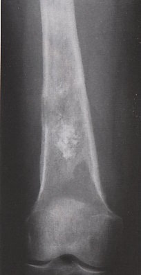
Is a primary malignant bone neoplasms are most often obtained. Occurred in the decade -two of life which is the active period of bone growth, with less than 5% occurred in children aged less than 10 years. Is highly malignant, rapidly metastasis to the lungs through the bloodstream.
Location: Learned in the metaphysis of long bones terut a ma on the distal femur, proximal tibia and proximal humerus.
Radiology:
Obtained three kinds of images an radiologists , namely:
A. picture osteolytic, where the destruction is the main process.
Two. picture osteoblastic, caused by a lot of bone tumor formation.
3. picture a mixture of the destruction and the formation of bone tumors.

Radiological image obtained a picture of osteolytic and osteoblatik, the MRI found a line destruction.
On MRI found the line due to the destruction and soft tissue extension of tumor cells.
Rapid growth resulted in the lifting of neoplasms poriosteum and reactive bone formed between the elevated periosteum and the bone-Ray padaX seen as Codman triangles. The combination of reactive bone and neoplastic bone formed along the blood vessels running radier than cortical bone in the direction of the image forming Sunbrust tumor.
Pathology: Overview histologinya vary. Criteria for the diagnosis of stromal sarcoma is gained with neoplastic osteoid formation of bone with a striking picture of anaplasia. Malignant cells to penetrate the cavity between the collection of osteoid.

Pathological picture of the discovery of stromal sarcoma and anaplasia.
Therapy: Patients with Osteosarcoma require operative therapy in the form of amputation.Amputation can be performed through the proximal bone tumor or through joints (disarticulation) proximal to the tumor. According to the investigation, the technique disarticulation amputation through the bone with no significant difference in the Survival Rate. In addition to operative treatment, it is also necessary in patients with osteosarcoma adjuvant therapy in the form of chemotherapy or radio therapy. Scientists say that at the time of diagnosis of osteosarcoma is made, it was thought most people already have micro-metastases in the lungs so that after amputation, although X-ray of lung metastases is still not visible, should be given chemotherapy to eradicate mikrometastase.
Prognosis: The prognosis Osteosarcoma is a bad beginning 5 years Survival Rate it just the range of 10-20%. This latter form of adjuvant therapy with an aggressive and intensive cytostatic given postoperatively then prabedah and Survival Rate to be better able to reach 60-70%. Thanks to therapy adjuvant therapy also has been reduced in recent amputation, now at cancer treatment centers are full, then the non-therapeutic amputation or Limb Salvage more frequent.
Parosteal Osteosarcoma / Juxtakortikal Osteosarcoma
Is a rare neoplasm derived. Obtained at the young age group, and most often on the posterior distal femur. The disorder is derived from the osteoblast cell periost and most often grows on the side of the bone. These neoplasms have a much different temperament osteosrakoma. The growth is slow when compared with metastatic osteosarcoma and also slow in the lungs. Therapy is wide excision, if necessary amputation. The prognosis is good, 5 years Survival Rate it more than 50%.
Sarcoma is rarely cause pain and pathological fractures.
Kondrosarkoma
Is a malignant tumor composed of cartilage cells ( cartilage ) that can grow spontaneously ( kondrosarkoma primer) or a malignant degeneration of benign lesions such as esteokondroma, enkondroma (secondary kondrosarkoma). Was found between 30-60 years of age. These neoplasms grow rather slowly and only very few complaints. Neoplasms are slow to give metastases.
Location: Mainly on the flat bones like the pelvis and the scapula, but can also be obtained in the long bones like the femur and humerus.
Clinical: patient complaint is the presence of the tumor becomes large gradually.


Onion-skinning Sunburst Codman's triangle


Type Expansif


Radiology: osteolytic lesion appears as a metaphysical middle of the bone with patches of calcified cartilage matrix derived from the process with cortical destruction, so the tumor can be extended to the surrounding soft tissue.

Pathological picture showed a lesion in the middle of the metaphysical with patches of calcification, the pathological picture found malignant cells in the middle lamellar.
Pathology: T ampak malignant cells between lamellar bone in the bone marrow that form cartilage.Mitosis picture is not so much.
Treatment: wide resection surgery, if necessary amputation. Adjuvant therapy such as radiotherapy, chemotherapy is not helping.
Fibrosarcoma
Connective tissue in the bone cavity is the origin of this malignant tumor, a malignant tumor of bone collagen matrix is formed but does not form osteoid or chondroid. Rarely available and is a malignant spindle cell noplasma. Incidence in women and men about the same. Fibrosarcoma may be connected with the presence of Paget's disease, fibrous dysplasia, giant cell tumor, chronic osteomyelitis. These neoplasms can also be a secondary neoplasm, as a result of radiation on benign bone tumor.
Clinic: Learned at the age of 20-60 years with complaints of severe pain in the lump that slowly enlarged. Pathological fractures may occur.
Location: On the long bones, most of the femur, tibia and humerus. Tumors obtained in the area of the lesion in the metaphysis and periosteal intramedular.
Radiology: It appears as a radiolucent lesion, as a result of bone destruction process cortikal, tumor boundary is unclear.
Pathology: tumor histological picture is seen as a malignant tumor in the presence of anaplasia, mitosis, there is a "herring bone pattern". Tumor cells inhibits the formation of collagen so that no osteoid surface / bone or cartilage. Microscopic picture is similar to soft tissue fibrosarcoma.
Therapy: D apat performed a radical excision and adjuvant radiation therapy.
Prognosis: T ergantung at the level of the tumor but 5 Years Survival Rate ± 25-30%, so it's better than kondrosarkoma or osteosarcoma.
Ewing's Sarcoma
Malignant tumors are rarely found. Also called "Small Round Blue Cell". Attacked the young age group, mostly under age 20 with a prevalence of approximately 80%. More to come on men.
Clinical: Patients complain of pain with a lump diseretai. The possibility exists that elevated body temperature, excessive sweating, lekositosis and increased erythrocyte sedimentation rate.
Location: In diafsisi long bones, most commonly in the femur, humerus, tibia, ulna and fibula, can also thin the bones.
Radiology: Looks process of bone destruction with no apparent limit. Reactive new bone formation by the periosteum in layers that can give you an idea Onion Skin or perpendicular to appear as Sunbrust.

Radiological picture of bone destruction with the boundary seem unclear, there is damage to the cortex MRI image.
On MRI showed cortical damage and disruption to the surrounding soft tissue.
Pathology: T erdapat picture of highly cellular, neoplastic infiltrates in the solid, round cells with clear cytoplasm "round blue cell tumor", and the presence of extensive necrosis as indicated by the occasional picture of Homer-Wright rossetes.
Pathological picture with noeplasma infiltrate, round blue cell tumors and Hommer-Wright rossetess.
Diagnosis: The clinical and radiological osteomeilitis similar to Ewing's sarcoma, osteosarcoma and metastatic neuroblastoma.
Therapy:
o Operation of the reaction area or amputation .
o Chemotherapy .
o Radiotherapy .
Prognosis: Poor. Mortality in the first years after diagnosis of ± 95%. Lately with kombinasai therapy radiotherapy, chemotherapy and surgery, the prognosis is better.
Retiku lo Sarcoma Bone
Malignant tumors are very similar picture mikroskopisnya Ewing's sarcoma, but have different clinical temperament. Acquired primarily in adulthood, particularly the long bones, pelvis and ribs.Grew more slowly than Ewing's sarcoma, resulting in less pain.
Locally, more destruksinya process, with the result of pathological fractures may occur.
Radiology: Similar to Ewing sarcoma .
Pathology: The microscopic wedding Ewing's sarcoma is distinguished by special staining. Tumor cells do not contain Kaposi reticulum glycogen (PAS negative), while Ewing's sarcoma cells contain glycogen (PAS positive).
Therapy: Radiosensitif. Of chemotherapy is also helpful. In some circumstances it may take action amputation.
Prognosis: Good. Survival Rate to 50%.
Myeloma multip el / plasma cell myeloma / Plasmasitoma
Malignant neoplasms of bone are often obtained due to the spread of osteolytic bone destruction. More to come in men over 40 years of age.
Clinical: Tumors cause pain. Can be found in patients with anemia, hypercalcemia, elevated body temperature, decreased body weight and feeling tired easily. No elevation in blood levels of gammaglobulin. Urine contains specific proteins that Bence Jones proteins, which can be found in approximately 50% of myeloma. Pathological fracture is a complication that often occurs because of the destruction process is fast with little reaction with reactive bone formation. The absence of reactive bone formation is often the cause of these tumors are not detected by bone scan. Diagnosis is confirmed by the examination of BMP (Bone Marrow puncture) in the sternum or iliac cyst which will see the abnormal plasma cells. Presence of Bence Jones protein in urine and elevation on the electro foresis gammaglobulin.
Location: In the elderly the bone marrow was still obtained vertebrae, pelvis and skull, and thoracic neoplasms are the most commonly found in the bones.
Radiology: punched out lesions resulting image, in the form of a rounded irregular osteolytic lesions.There is no periosteal reaction. Erosion starts from intramedular up to the cortex.
Pathology: T erdapat plasma cells, osteolytic lesions caused by increased osteoclast resorbsi cytokines influenced by plasma cells.
Therapy: Radiotherapy and chemotherapy, or bone marrow transplants performed in pathological fractures of the limb bones do internal fixation.
Prognosis: Poor, 5 Years Survival Rate ± 10%. Patients usually die 2 years after diagnosis.
Angiosarcoma
A malignant tumor dalah rare , a histologist is characterized by the formation of an irregular vascular anastomosis is covered by one / more endothelial cells are atypical and unusual picture of the immature cells is accompanied by a less dense mass of undifferentiated (anaplastic) . Lesions in this disorder are multiple lesions in one bone, then very quickly going to the surrounding bone metastasis to the lung or lungs .
Lympangioma fast growth can result in extensive bone destruction (massive osteolysis) and gives an overview of X-Ray dissapearing bone or bone phantom.
Location: T erutama the long bone on bone diafisis.
Clinical: N Yeri heavy on the diafisis tulong mainly lower limb bones.
Radiology: T erdapat picture of lesions on the mid-diafisis ipsilateral or contralateral bone and on the part of the first lesion. Thoracic CT scan showed nodules in the lungs, if carried out will be enforced biopsy diagnosis of high-grade angiosarcoma.
Therapy: A ika is found distant metastases, surgical treatment is no longer the choice of therapy, only therapy to prevent pathological fractures. With radiation and adjuvant therapy can be given.
Prognosis: If the tumor did not respond to radiation, it is possible patients may only last up to 1 year after diagnosis.
Giant Cell Tumor / Tumor Cells Giant / Osteoklastoma
The origin of bone tumors is still controversial, some have argued this tumor derived from connective tissue, the other opinion says these tumor cell origin of osteoclasts, but there is also the opinion of this tumor from unknown origin. These tumors have a nature and a tendency to become malignant and aggressive so it is categorized as a malignant tumor .
Giant cell tumor ranked second (17.5%) of all malignant bone tumor, mainly found at the age of 20-40 years and rare below the age of 20 years and more frequently in women than men.
Location: Learned in the epiphysis of long bones that may extend towards the metaphysical. The most common is the proximal tibia, distal femur and distal radius. Can also be found in the pelvis and sacrum.
Clinical: K eluhan increasing pain and swelling in the bone lesions , especially in the knee and joint effusions may be found in the joints and movement disorders.
Radiology: Looks osteolytic areas in the epiphyseal with clear boundaries and gives the impression of a picture multilokuler soap bubble. Cortical thinning occurs.

Osteolytic radiological picture seen in the epiphyseal area with a soap bubble, there is a pathological picture datia cells and tumor cell mitosis.
Pathology: P No dosage curettage results, a vascular stroma with many cells datia / giant cell.Mitosis was found with ease on 4x and 10x magnification. Some places are fibrihistiocit cell and cell xantoma.
Treatment: Surgery followed curettage with bone graft or bone filling cement. And some of adjuvant therapy with phenol, insertion PMMA (polymethylmetacrylate), cryotherapy after curetase.
On some things can be done tumor resection, wide excision is accompanied by reconstruction measures. Sometimes require amputation.
Kordoma
M erupakan malignant tumor originating from the rest notokordal, often found in young adults with the highest prevalence at ages 50 to 70 years. Comparison of the incidence in men and women is 2:1.
Location: T umor is growing progressively in the sacrum and coccyx (50%). Kordoma can also be found on the spine and will provide lower back pain. In the event of the sacrum, it may cause urethral obstruction kordoma / rectum and may occur in advanced neurological symptoms. There are also reports that these tumors can occur in the bones of the head.
Clinical: G ejala kinis depending on where it happens, sometimes there are complaints of pain, disorders of the urogenital system, if headaches occur in the bones of the head.
Radiologist: a plain radiological picture can be found is a radiolucent image in the sacral region that showed a lesion with bone destruction. There is a focal calcification. With CT-scan and MRI can be seen spread and tumor enlargement intrapelvik, calcified epidural.
Pathology: The tumor is characterized by the arrangement of lobuler and septal tissue formed from cells that contain many vacuoles (fisaliporus cells) with a mucoid intercellular material to be mucus.
Treatment: Treatment can be done in the form of local resection. Tumors usually infiltrate the surrounding soft tissue metastases but rarely held. Good prognosis of this disorder is rare in recurrent. But be aware of the anatomical location not to interfere with sexual function.
Adamantinoma
M erupakan tu mor rare malignant bone, especially regarding diafisis tibia (90%) and are often found at the age of 20-25 years.
These tumors included in the tumor of low grade and metastasize slowly.
These histological tumor as with adamantinoma of the jaw (ameloblastoma of the jaw).
Location: T erutama diafisis lies in the tibia, also in the jawbone (mandible).
Clinical: N Yeri blunted in chronic bone, soft tissue pain around the surrounding bone pain.
Radiological: radiological picture looks a typical bubble defect in the anterior tibial cortex. May also be found thickening of the surrounding bone. Radiolucent lesion around the bone sclerosis, and there are also small lesions with a similar picture. With a CT-scan can be seen in the spread of tumors to spread beyond the medulla or periosteum.
Radiological picture looks radiolucent lesion with a ge l umbung typical.
Pathology: The histological examination found the island that resembles epithelial cells with intercellular selspindle network. There is also a picture of a fibrous stroma cells are composed of nests of basal cell contains a dark picture.
Pathological picture of the island of cells found in spindle cell nuclei .
Treatment: The treatment of adamantinoma Basically a wide local excision with taking parts of the surrounding normal bone. CT-scan examination is necessary before surgery to see inside tumor penetration. When the lesion extends to the endosteal surface of the excision should be performed on all segments and the remaining empty space is filled with bone graft that bervaskularisasi.
Secondary bone tumors
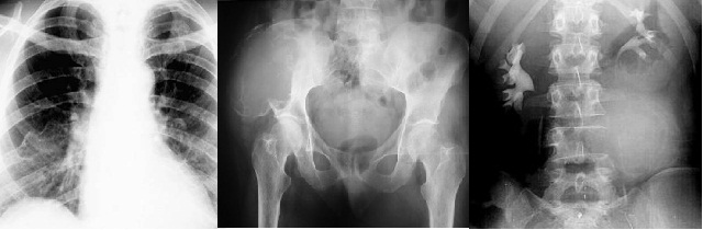
Is a type of malignant bone tumor that is often obtainable. Possible bone tumor is a metastatic tumor should always difikirkan, in patients who are elderly. At the age of consent / advanced type of malignancy is often metastatic to bone are breast carcinoma, lung, stomach ,kidney, colon, prostate and thyroid.
Being of children is neuroblastoma. Sufferers who died of carcinoma, the post-mortem examination was at least a quarter showed signs of bone metastases. Daughter cells reach the bone by spread through the blood, lymph channels or by direct extension. Bone marrow is a fertile place for growth in daughter cells spread, thereby vertebrae, pelvis, ribs and the proximal long bones are the most often seized by the daughter cells spread.
In patients with bone malignancy metastasik possibility, then the examination should be performed on all the bones such as the bone surveys or bone scans. The most prominent complaint of patients is pain. The pain can be caused by pathologic fracture.
In some circumstances it ditulang metastatic lesions are first discovered and diagnosed, where the results of microscopic examination showed a type of metastatic bone neoplasms is sometimes difficult to determine tissue of origin, so it should look carefully than the primary tumor site.
Radiology: In general, metastatic tumors will result in osteolytic picture, is the apparent picture of metastatic prostate Ca osteoblatik / osteosclerosis. Elevated calcium levels due to the release of calcium into the blood due to osteoblastic resorption process in the bones. The presence of reactive bone formation is characterized by increased levels of alkaline phosphatase. In the metastasis of prostate Ca, elevated acid phosphatase levels.
Treatment: Treatment is palliative, because the patient was in an advanced stage. Therapy is aimed at primary carcinoma type that may include radiotherapy, chemotherapy or hormone therapy. In terms of surgical therapy is of pathological fractures that may require external or internal fixation, so that patients can be immobilized without feeling pain. If you need help to do internal fixation of the bones of the limb before the bone is fractured, so the new process is expected to fracture when the bone is left goes on (impending fracture).
DIAGNOSIS
To establish the diagnosis of bone tumors need a few things, namely:
Anamnesis
History is important to know the history of the disorder or previous trauma. It should also be asked whether there is a family history of similar illnesses such as diafisial aklasia that is hereditary.
Some things to note in history are:
A. Age
Age of patients is very important to know because a lot of bone tumors that have a quirk in the age of, for example Osteogenik sarcoma found in children up before the young adults, kondrosarkoma at the age of 40 years, giant cell tumor is rare under the age of 20 years.
2. Old and development (progressive) tumor
Benign tumors usually grow slowly and if there is rapid development in a short time or a benign tumor that has suddenly become big it is necessary to suspect the presence of malignancy.
3. Pain
Pain is the main complaint in malignant tumors. The pain showed signs of rapid expansion and the suppression of tumor into surrounding tissue, bleeding or degeneration.
4. Swelling
Sometimes patients complain of a swelling, which arose gradually within a period of time or suddenly.
Clinical Examination
Things that are important in clinical examination are:
A. Location
Several types of tumors have a classic location and places a particular predilection as in epiphyseal regions, metaphysical bone, or attack certain bones such as osteoma in the skull, in the metaphysical Osteogenik sarcoma, osteoblastoma in the vertebral region.
2. Large, shape, boundaries and nature of the tumor
Tumors are less likely a benign tumor, whereas tumors that most likely is a malignant tumor. Is also important to note the form of tumor is accompanied by dilation of blood vessels or ulcers that are characteristic of a malignant tumor. Signs of joint effusion may be found in the tumor adjacent to the joints.
3. joint movement disorders
In the large tumors around the joint will give interference with joint movement.
4. muscle spasm and stiffness of the spine
If there is tumor in the spinal area, either benign or malignant, can provide a spasm / stiffness of the muscles of the spine.
Five. pathological fracture
Some malignant tumors may provide a pathological fracture complications due to bone fragility occurs so that the patient will present with symptoms of fracture.
Neurological examination
If there are symptoms in patients with neurological disorders, the neurologic examination should be done carefully to determine whether this disorder arises because of the emphasis on certain nerve tumor.
Radiological examination
Is one of the most important examination in the diagnosis of bone tumors. Performed locally on plain or photo lesion site surveys throughout the bone (bone survey) if the suspected presence of a tumor that is metastatic or primary tumor that can affect several parts of the bone.
Plain bone can give you an idea:
• Location of tumors: epiphysis, metaphysis, diafisis or in certain organs.
• Characteristically solitary or multiple.
• Type of the affected bone.
• Limit firm tumor / no, contain calcifications / no.
• cystic or solid form.
Other radiological examination to do, namely:
ü Radionukleida scanning , the examination is usually used on small lesions such as osteoma.
ü CT scans, can provide information about the existence of intraosseous tumor whether or ekstraoseus.
ü MRI, can provide information on whether the tumor is located in the bone or not, whether the tumor expands into the joints or soft tissues.
DESCRIPTION OF CONVENTIONAL Radiography
Picture of the lesion
- Destruction of bone
- periosteal reaction
- Matrix of tumor
- Expansion of lesions
- The involvement of soft tissue
Type of destruction there are 3 kinds:
Geographic
Moth-eaten
Permeative
Geographic Bone Destruction
Limit firm
Indications à less aggressive, slower growth, benign
narrow transition zone
Geographic lesions
Examples
Examples
Non-ossifying fibroma
Chondromyxoid fibroma
Eosinophilic granuloma
Moth-eaten destruction
Limit uneven, ragged
Indications à
growing faster than the geographic
Perhaps the malignancy
Moth-eaten

Examples
Myeloma
Metastases
Lymphoma
Ewing'S sarcoma
Permeative
no clear boundary with "worm-holes" multiple
The spread through the bone marrow space
The transition zone width
Indication
Malignant aggressive (malignant, aggressive)
Permeative
Examples

Lymphoma, leukemia
Ewing'S Sarcoma
Myeloma
Osteomyelitis
Neuroblastoma
Type of destruction


Periosteal reaction
Benign
o None
o Solid
Malignancy
Lamellated or onion-skin
Sunburst
Codman's triangle
Type of periosteal reaction

Type of periosteal reaction
Benign
None

Non-ossifying fibroma
Solid
Chronic osteomyelitis

Onion-skinning Sunburst Codman's triangle
|


Tumor matrix
osteoblastic
o Fluffy, cotton-like or cloud-like densities
§ Osteosarcoma
Cartilaginous
o Comma-shaped, punctate, annular, popcorn-like
§ Enchondroma, chondrosarcoma, chondromyxoid fibroma
Tumor matrix

Osteosarcoma Chondrosarcoma
Type Expansif

Brown fibrous dysplasia Enchondroma Lymphoma Tumors
Soft tissue involvement
Indication
Common malignancies
Asmass soft tissues that firmly
benign lesion with soft tissue involvement
Osteomyelitis

Osteosarcoma
Multiple lesions
More often benign
Multiple malignant lesions
Metastatic

Lymphoma
Mets from Ca of Prostate
Multiple Myeloma


Osteosarcoma

Multiple Myeloma Osteokondroma


Laboratory
Blood, include: LED examination, hemoglobin, serum alkaline phosphatase, serum protein electrophoresis, serum acid phosphatase that provide diagnostic value in malignant bone tumors.
Urine , critical examination is the examination of protein BenceJones .
Biopsy examination
The goal is to obtain sufficient material for histological examination, to help establish a diagnosis, and tumor staging (determining malignancy).The timing of the biopsy is very important because it can affect the results of radiological examinations are used in staging. If a CT-scan was made after a biopsy is performed, it will be visible bleeding in soft tissue that gives the impression of a picture of a malignancy in soft tissue.
There are two methods of biopsy, namely:
o Biopsy closed, using FNAB (Fine Needle Aspiration Biopsy) to perform sitodiagnosis. Is one way to do a biopsy on the tumor diagnosis.
Advantages of FNAB were:
-No need of care.
-The risk of minor complications.
-Prevent the spread of tumors.
- Quickly get results.
o an open biopsy, the biopsy method through operative measures. The advantage is that it can take a larger network for histological examination and inspection ultramikroskopik, reduce errors and reduce the tendency of making the network diagnostic differences in benign and malignant tumors such as between enkondroma and kondrosarkoma, osteoblastoma and osteosarcoma. Open biopsy should not be done when it may cause difficulties in subsequent surgical procedures, such as the resection en-bloc .
DIAGNOSIS
Needs to be done with extreme caution because of other abnormalities such as infection, osifikans myositis, hematoma may provide the clinical and radiological picture resembling a malignant tumor of bone. Abnormalities that may provide clinical and radiological picture resembling a tumor, among others:
A. subperiosteal hematoma or soft tissue that would provide a lump that ache.
2. Osteomyelitis can give symptoms such as malignant tumor malignant osteosarcoma or Ewing's tumor.
3. fracture stress; this will provide an overview of the symptoms of pain and fracture.
4. myositis osifikans.
5. arthritis gout.
MANAGEMENT
In the malignant tumors required the cooperation and consultation between oncology surgeons, orthopedic surgeons, radiologists, pathologists as well as prosthetics and rehabilitation experts.
v would Suspicious malignant tumor
If a lesion in the primary tumor as a malignancy is suspected then the patient should be treated for a complete examination, blood tests, lung images, either with plain imaging and CT-scan and biopsy of the tumor.
Operation
Excision of the tumor by surgery can be performed by several techniques, namely:
Ø intralesional or intrakapsuler
This technique is performed by excision / curettage of the tumor, is not recommended in malignant tumor and the tumor is usually performed on low grade tumor , such as giant cell tumor.
Ø marginal excision
Tumor is spending outside of the capsule. This technique is mainly done on the tumor is benign or malignant tumor types of low grade malignancy .
Ø Wide excision (excision of the en-bloc )
Tumor removed intact tissue around the tumor with a form of pseudo-capsule or tissue outside the tumor reacts. Actions performed on the wide excision of malignant tumor and is usually combined with chemotherapy or radiotherapy in the pre / post surgery.
Ø Operating radical
Performed as in the wide excision plus expenses and all the bones and joints and tissues as an integral part. This method is usually a limb amputation on it and with the joint expenditure on it.
With proper staging and chemotherapy to control the spread of tumor, amputation of action can be avoided with a technique called limb-sparing surgery (limb saving procedure) is a wide excision is accompanied by the replacement limb by using a bone graft or protesis tailored to members The motion specially made individually.
Radiotherapy
With high-energy radiation is a way for the eradication of malignant tumors and may also radiosensitif as initial treatment prior to surgery performed. The combination of radiotherapy can also be given together with chemotherapy. Radiotherapy is performed on the circumstances of the in-Operable example of metastasis or local circumstances do not allow for surgery.
Chemotherapy
An additional treatment on malignant tumors of bone and soft tissue. The drugs used were methotrexate, adriamycin, cyclophosphamide, vincristine, sisplatinum. Chemotherapy is usually performed on pre / post surgery.
CHAPTER III
CONCLUSION
Tumor of bone is a bone disorder that is neoplastic. In other terms "Tumor Bones" is an abnormal growth of bone that can be benign or malignant.
Of all primary bone tumors; 65.8% 34.2% benign and malignant. This means that out of every three have a bone tumor that is malignant. Comparison of the incidence of bone tumors in men and women are equal.
Classification based on origin of cell neoplasms of bone , namely: primary (Osteogenik, kondrogenik, fibrogenik, mielogenik, tumor vascular origin, the other bone tumor) and secondary /metastatic (t umor derived from other organs that spread to the bone ).
To establish the diagnosis of bone tumors need a few things, namely: anamnesis, clinical examination, neurological examination, radiological examination, laboratory tests, and biopsy examination.
Method of treatment on bone neoplasms such as by surgery, radiotherapy, and chemotherapy. There are several surgical techniques, namely: intralesional or intrakapsuler, marginal excision, wide excision and radical surgery.
REFERENCES
· Doherty, Gerard M. Current Surgical Diagnosis & Treatment 12th Chapter 42 page 1201 to 1205. Mc Graw Hill
· Rasjad C. Introduction of Orthopaedic Surgery . New York: Yarsif Watampone. 2007 , p.275-315.
· Reksoprodjo S. Lecture set of Surgery . New York: Binarupa script. 1995 , p. 331-340.
· Sjamsuhidajat R, Wim de Jong. Textbook of Surgery , 2nd edition. New York: EGC. 2005, p. 934-939.
· Suyadi A. Bone Malignancy . New York: FK UII Surgery. 2009, p. 1-11.
No comments:
Post a Comment