"A Man can't make a mistake can't make anything"

Monday 30 April 2012

STANDARD TREATMENT OF PATIENTS HIV / AIDS (acquired immune deficiency syndrome), WHEN HAVE SURGERY. STANDAR PENANGANAN TERHADAP PENDERITA HIV / AIDS, KETIKA MENGALAMI PEMBEDAHAN (DEFINITION, SIGN, SYMPTOMS, DIAGNOSIS, PROGNOSIS AND MANAGEMENT)


STANDARD TREATMENT OF PATIENTS HIV / AIDS, WHEN HAVE SURGERY

CHAPTER I
INTRODUCTION

HIV infection is a viral infection that progressively destroys white blood cells and causes AIDS (Acquired Immunodeficiency Syndrome). (10) AIDS was first discovered in 1981. (1)
Transmission of HIV infection from mother to child is the main cause of HIV infection in children under 15 years of age. Since HIV became pandemic in the world, an estimated 5.1 million children globally are infected with HIV. Most of these patients infected through transmission from mother to child. Each year an estimated more than 800,000 infants become infected with HIV through transmission from mother to child. And followed by the approximately 610 000 child deaths from the virus. (10)
            In Indonesia, according to the Directorate General of Health Department of PPM and PL recorded 3568 cases of HIV / AIDS at the end of December 2002. There are 20 children with HIV infection who are infected mother. Research conducted Pelita Science Foundation and The Faculty of medicine obstetrics / RSCM during the years 1999-2001 do a check on 558 pregnant women in poor areas in Jakarta, showed as many as 16 people (2.86%) suffering from HIV infection. (10)
Women are contracting HIV infection through heterosexual relations with an infected partner or through the use of drugs, increasing HIV infection in children is due to the result of transmission during the perinatal period (pregnancy, during and after childbirth). Perinatal HIV transmission is a major cause of pediatric AIDS mother - mothers that transmit HIV are usually good and has a number of CD 4 T lymphocytes are normal and do not know if they are infected with HIV. More than 90% of AIDS in children were reported in 1994 occurred because of transmission from mother to child. (10)
 Transmission to the infant can occur during pregnancy, delivery or postnatal through breast milk. The incidence of mother to child transmission is estimated at 20% - 30%. HIV transmission to the fetus if no intervention reported to range between 155-45%. (10)
The risk of transmission in developing countries around 21% - 43%, higher than the risk of transmission in developed countries about 14% -26%. Transmission can occurs during pregnancy, intrapartum, and postpartum. The risk of transmission of most infections occur during labor by 18%, in the content of 6% and 4% after delivery.


Thursday 26 April 2012

MY HEAD ENLARGED AND MY SUN SET EYES : HYDROCEPHALUS (definition, anatomy, diagnosis, sign, symptom, and management)


CHAPTER I
INTRODUCTION

Hydrocephalus comes from hydro meaning water and chepalon which means head. Hydrocephalus is a buildup of cerebrospinal fluid (CSS) is actively causing dilatation of the ventricular system of the brain where there is excessive accumulation of CSF in one or more of the ventricles or subarachnoid space. This situation is caused because there is imbalance between production and absorption of the CSS. If the excessive accumulation of CSF occurs on the cerebral hemispheres, a condition called subdural higroma or subdural fluid collection. In the case of excessive fluid accumulation occurs in the ventricular system, a situation known as hydrocephalus internal.Selain that some intracranial lesions causing elevation of ICT, but not until the cause of hydrocephalus. CSS is not equivalent to raising the volume with hydrocephalus; it also occurs in cerebral atrophy.


Hydrocephalus as a clinical entity distinguished by three factors: a). Intraventrikuler pressure elevation, b). Addition of CSS volume, c). Dilatation of the cavity CSS. Overall, the incidence of hydrocephalus is estimated close to 1: 1000. while the incidence of congenital hydrocephalus varies for each of the different populations. BL Hershey said most of hydrocephalus in children is congenital is usually seen in infancy. If hydrocephalus appears after age 6 months is usually not because of congenital. Mujahid Anwar et al get 40-50% of infants with intraventricular hemorrhage grade 3 and 4 had hydrocephalus. Pongsakdi Visudiphan et al in a study 36 of 49 children with TB meningitis had hydrocephalus, with a second note 8 children with obstructive hydrocephalus and 26 children with communicating hydrocephalus. Hydrocephalus that occurs as a complication of bacterial meningitis can be found at any age, but more often in infants than children.

Tuesday 24 April 2012

Basic skills of medical personnel: CPR / Cardiopulmonary Resuscitation


CHAPTER I
INTRODUCTION
1.1 Background
Cardiac Pulmonary Resuscitation is very important in the life of the medical world, so that every doctor should be able to perform Cardiac Pulmonary Resuscitation. Well as other paramedics. In fact, any layperson who works in a place where a lot of people (the crowd) or the place where a lot of work to contain the risk of work should be able to be trained in Cardiac Pulmonary Resuscitation is a simple to a more competent medical officer. B-gels or in the Indonesian language known as the Emergency First Aid (PPGD) is the set of the first attempts to do in emergency conditions in order to save patients from death. Abroad, PPGD is actually taught in many ordinary people or ordinary people special, but it seems this is still very little known by the people of Indonesia.

1.2 Key Principles
 PPGD is the main principle of saving the patient from death in emergency conditions. Later in the PPGD philosophy is "Saving Time is Life Saving", in the sense that all actions taken during emergency conditions to be truly effective and efficient, because the patient's condition may have lost their lives in just minutes (stopping breathing for 2 -3 minutes can cause death).

1.3 Basic Steps
The basic steps in PPGD known by the acronym ABCD (Airway - Breathing - Circulation - Disability). These four points are the points that should be kept in the response of patients in emergency conditions.

1.4 Pulmonary Resuscitation heart
Stop the breath because:
- Airway Obstruction
- Foreign bodies
- Aspiration
- The tongue falls down
- Pipe folded endotracheal
- Blocked tracheal cannula
- Acute Abnormalities of the glottis and surrounding
- Depression of central respiratory
- Drugs
- Intoxication
- High pCO2
- Low pO 2
- After the cardiac arrest
- Brain tumors
- Sunset
Peripheral:
- Drug muscle paralytic
- Myasthenia gravis
- Poliomyelitis

For the Stop Heart
1. Cardiovascular (heart disease siskemik, IMA, pulmonary embolism and conduction system fibrosis)
2. Acute oxygen deficiency (stopping breathing, and obstruction due to foreign body secretions)
3. Overdose of drugs (digitalis and adrenaline)
4. Acid-base disturbances / electrolyte (K increases or decreases, increased Mg, Ca increases, acidosis)
5. Accidents (electric shock, drowning)
6. Vagal reflex
7. Anesthesia and surgery
8. Medical diagnostic and therapeutic measures
9. Shock, cardiac arrest can be accompanied by electrical phenomena.
Adam Stokes syndrome
Condition caused by high degree AV block heart is characterized by episodic bradycardia or asystole that resulted in the attack who are not aware of themselves suddenly with / without a seizure action.
- Artificial Circulation
- External cardiac massage
Indications CPR: Stop or stopping breathing and circulation.
Contraindications: Cardiac arrest, rib fractures, thoracic fractures, cardiac tamponade, pneumothorax, severe emphysema, Cardiac Arrest over 5-6 minutes.

Monday 23 April 2012

BENIGN OR CANCER (MALIGNANT) PAROTID GLAND TUMOR (definition, sign, symptoms, diagnosis, prognosis, and management

Parotid gland


Anatomy of parotid gland
Parotid gland is the salivary glands are paired, numbered 2. Parotid gland is the largest of the salivary glands. Each weighing an average of 25 grams and irregular shape, berlobus, color between green and yellow (Yellowish) located below the external acoustic meatus between the mandible and the muscles sternokleidomastoideus.4

Parotid gland varied shape, when viewed from the lateral triangular 50%, 30% of the top and bottom round. Parotid gland is usually shaped like an inverted pyramid with a surface-surface as follows: superior surface of the small, superficial, anteromedial, and posteromedial. Konkav on the superior surface forms associated with the cartilage of the external acoustic meatus and the posterior part of the temporomandibular joint. Here auriculotemporal nerve supplies the parotid gland. Superficial surface covered by skin and superficial fascia containing a branch of the facial nerve aurikuler, superficial parotid lymph nodes, and the lower limit of platisma.4
The anterior part of the gland adjacent to the posterior edge of mandibular ramus and slightly coat the posterior edge of masseter muscular. Posterior part of the gland is surrounded by the ear, mastoid processus, and the anterior edge of the muscular stemokleidomastoideus. The inside of the medial lobe extends into the cavity parafaring, limited by the processus stilomandibular stiloideus and ligament, muscular digastrikus, and carotid sheath. In the anterior lobe is located adjacent to the medial ptetygoideus. Lateral parts covered only by skin and subcutaneous fat tissue. Connective tissue and fat tissue from the fascia of the neck in this gland wraps. Parotid gland is closely linked to important structures around the internal jugular vein and its branches, the external carotid artery and its branches, lymph glands, auriculotemporalis of nerve branches and nerve trigerninus fasialis.4

Thursday 19 April 2012

TRUE EMERGENCY : HEAD INJURY, WITH EPIDURAL HEMATOMA /cedera kepala, dengan epIidural hematom. (sign, symptoms, etiology, diagnosis, management)


Epidural hematoma is a type of intracranial bleeding most often occurs due to fracture of the skull. Olek cover the brain in the skull bones are rigid and hard. The brain is also surrounded by something that is useful as a wrapper which is called the dura. Its function is to protect the brain, blocking the venous sinuses, and form the periosteum tabula interna.

I. INTRODUCTION
Epidural hematoma is a type of intracranial bleeding most often occurs due to fracture of the skull. By cover the brain in the skull bones are rigid and hard. The brain is also surrounded by something that is useful as a wrapper which is called the dura. Its function is to protect the brain, blocking the venous sinuses, and form the periosteum tabula interna .. When one gets a great impact on the head is likely to form a hole, the movement of the brain may cause abrasion or laceration of blood vessels surrounding the brain and dura, when a blood vessel had torn the blood will accumulate in the space between the dura and the skull, the state inlah are often known as an epidural hematoma.
Epidural hematoma as a state of emergency and the neurologist who is usually associated with a linear fracture that decides the larger arteries, causing bleeding. Venous epidural hematoma associated with vein laceration and progress gradually. Arterial hematoma occurred in the middle meningeal artery that lies beneath the temporal bone. Bleeding into the epidural space, so if there is bleeding artery hematoma will quickly occur.

II. INCIDENCE AND EPIDEMIOLOGY
In the United States, 2% of cases of head trauma resulting in epidural hematoma and about 10% resulting in a coma. Internationally frequency of occurrence of epidural hematoma is similar to the incidence in the United States who are at risk of edh . are parents who have problems walking and frequent falls.
60% of patients with epidural hematoma is under the age of 20 years, and rarely occurs in less than 2 years of age and over 60 years. The increased mortality in patients aged less than 5 years and more than 55 years. Occurs more frequently in males than in females with a ratio of 4:1.
Types:
1. Acute epidural hematoma (58%) of arterial bleeding
2. Subacute hematoma (31%)
3. Cronic hematoma (11%) bleeding from vena

III. Etiology
Epidural hematomas can happen to anyone and any age, some circumstances that can lead to epidural hematoma is such a collision on the head on motorcycle accident. Epidural hematoma caused by head trauma, which is usually associated with fracture of the skull and laceration of blood vessels.

IV. ANATOMY OF THE BRAIN
The brain is protected from injury by the hair, skin and bones and wrap it, without this protection, the soft brain that makes us like it is, it would be easier to injury and damage. In addition, once a neuron is damaged, can not be repaired anymore. Head injuries can lead to big disaster for someone. Most of the problem is a direct result of head injuries. These effects should be avoided and immediately found from the medical team to avoid a series of events that lead to mental and physical disorders and even death.
Right at the top of the skull lies aponeurotika galea, a fibrous tissue, dense can move freely, which memebantu absorb the force of external trauma. In between the skin and galea there is a layer of fat and membrane layers in the vessel- large. If the tear is difficult to hold a vessel vasoconstriction and can cause significant blood loss in patients with lacerations on the scalp. Just below the galea are subaponeurotik space containing veins and diploika emisaria. These vessels can  infection of the scalp until deep into the skull, which clearly shows how important cleansing and debridement of the scalp galea carefully when torn.
In adults, the skull is a tough room that is not possible intracranial extension. Bone actually consists of two walls or a tabula separated by a hollow bone. Outer wall in  tabula externa, and the inner wall is called tabula interna. Structure and thus allows a force greater isolation, with a lighter weight. tabula interna contains grooves  anterior meningeal artery, the media, and p0osterior. If the fracture of the skull caused tear one of these artery-artery, which in  arterial bleeding, which accumulated in the epidural space, can  fatal consequences unless it is found and treated promptly.
Other protective lining of the brain are the meninges. The third layer of the meninges is the dura mater, arachnoid, and pia mater:
1. Cranial dura mater, the outer layer is thick and strong. Consists of two layers:
- Endosteal layer (periosteal) formed by the outer periosteum wrapped in calvari
- The inner meningeal layer is a strong fibrous membrane that goes on in the foramen magnum with the spinal dura mater that surrounds the spinal cord
2. Arachnoidea mater cranial, intermediate layer that resembles a spider's web
3. Cranial pia mater, the innermost layer of which contains many fine blood vessels.

Figure 1. Anatomy of the head

KODE ETIK KEDOKTERAN INDONESIA ( KODEKI ) DAN PEDOMAN PELAKSANAAN. MEDICAL ETHICS CODE OF INDONESIA AND IMPLEMENTATION GUIDELINES

Bersama ini saya downloadkan KODEKI dari website IDI  mudah-mudahkan bisa bermanfaat bagi sejawat sekalian dalam memahami KODEKI KEDOKTERAN.


KODE ETIK KEDOKTERAN INDONESIA
DAN
PEDOMAN PELAKSANAAN
KODE ETIK KEDOKTERAN INDONESIA

MAJELIS KEHORMATAN ETIK KEDOKTERAN INDONESIA
(MKEK)
IKATAN DOKTER INDONESIA (IDI )

Jl. Dr. Samratulangi No. 29
Telp. 3150679 – 3900277; Fax 3900473
Jakarta 10350


PEDOMAN PELAKSANAAN KODE ETIK KEDOKTERAN INDONESIA


PENGANTAR

Muktamar IDI XXI Tahun 1991 di Yogyakarta menetapkan Kode Etik Kedokteran Indonesia dan pedoman pelaksanaan KODEKI yang isinya merupakan hasil seminar tertulis penyempurnaan Kode Etik Kedokteran Indonesia yang diselenggarakan oleh Majelis Kehormatan Etik Kedokteran (MKEK) Pusat dan beberapa penyempurnaan oleh Muktamar. Dengan demikian KODEKI yang semula terdiri dari Pasal-Pasal dan penjelasannya maka sesuai dengan ketetapan Muktamar XXI disusun menjadi:
1. KODEKI
    Berisi pasal dan penjelasan ringkas.
2. Pedoman Pelaksanaan Kode Etik Kedokteran Indonesia.
     Berisi penjelasan dan petunjuk serta contoh pelaksanaan KODEKI.
   Pada hakikatnya kedua buku tersebut bukan dua hal terpisah, namun harus dilihat sebagai satu kesatuan.


KEMAMPUAN DASAR MEDIS : RESUSITASI JANTUNG PARU / CARDIO PULMONARY RESUSCITATION ( CPR)


BAB I
PENDAHULUAN

1.1 Latar Belakang
Resusitasi Jantung Paru sangat penting dalam kehidupan dunia medis, sehingga setiap dokter harus bisa melakukan Resusitasi Jantung Paru. Juga petugas paramedis lainnya. Malah orang awam pun yang bekerja di tempat tempat yang banyak orang (keramaian) atau tempat tempat pekerjaan yang banyak yang mengandung resiko kerjaan harus bisa untuk dilatih Resusitasi Jantung Paru yang sederhana sampai petugas medis yang lebih berwenang datang. B-GELS atau dalam bahasa Indonesia dikenal dengan Pertolongan Pertama Pada Gawat Darurat (PPGD) adalah serangkaian usaha-usaha pertama yang dapat dilakukan pada kondisi gawat darurat dalam rangka menyelamatkan pasien dari kematian. Di luar negeri, PPGD ini sebenarnya sudah banyak diajarkan pada orang-orang awam atau orang-orang awam khusus, namun sepertinya hal ini masih sangat jarang diketahui oleh masyarakat Indonesia. 

1.2 Prinsip Utama
 Prinsip Utama PPGD adalah menyelamatkan pasien dari kematian pada kondisi gawat darurat. Kemudian filosofi dalam PPGD adalah “Time Saving is Life Saving”, dalam artian bahwa seluruh tindakan yang dilakukan pada saat kondisi gawat darurat haruslah benar-benar efektif dan efisien, karena pada kondisi tersebut pasien dapat kehilangan nyawa dalam hitungan menit saja ( henti nafas selama 2-3 menit dapat mengakibatkan kematian). 
1.3 Langkah-langkah Dasar 
Langkah-langkah dasar dalam PPGD dikenal dengan singkatan A-B-C-D ( Airway - Breathing – Circulation – Disability ). Keempat poin tersebut adalah poin-poin yang harus sangat diperhatikan dalam penanggulangan pasien dalam kondisi gawat darurat.

1.4 Resusitasi jantung Paru
Sebab Henti nafas :
- Sumbatan jalan nafas
- Benda asing
- Aspirasi
- Lidah jatuh kebawah
- Pipa endotrakeal terlipat
- Kanul trakeal tersumbat
- Kelainan akut glottis dan sekitarnya
- Depresi pernafasan sentral
- Obat
- Intoksikasi
- pCO2 tinggi
- pO2 rendah
- Setelah henti jantung
- Tumor otak
- Tenggelam 
Perifer  :
-    Obat pelumpuh otot
- Miastenia gravis
- Poliomielitis

Tuesday 17 April 2012

ORAL-ONCOLOGY SURGERY SERIES : ORAL CAVITY CANCER / kanker rongga mulut ( definition, sign, symptoms, etiology, diagnosis and management)


CHAPTER I
INTRODUCTION

Diseases of the oral soft tissues has been a serious concern by experts, especially with the rising cases of deaths caused by cancer in the oral cavity particularly in countries that are developing.
Oral cavity cancer is approximately 5% of all malignancies occurring in men and 2% in women (Lynch, 1994). It has been reported that oral cancer is the leading cancers in India especially in Kerala where the incident was reported at an average height, about 20% of all cancers (Balaram and Meenattoor, 1996).
Although there are developments in the diagnosis and therapy, abnormalities and mortality resulting from oral cancer is still high and has long been a problem in the world. Several reasons are put forward for this is mainly due to the lack of early detection and identification of high risk groups, as well as failure to control the primary lesion and cervical lymph node metastasis (Lynch, 1994; and Meenattoor Balaram, 1996).
To overcome the problems caused by oral cancer, WHO has made instructions to control oral cancer, especially for countries that are developing. Control is based on primary prevention measures where the main principle to reduce and prevent exposure to substances that are carcinogens. The second approach is through the implementation of secondary prevention, ie early detection of cancerous lesions and precancerous oral cavity (Subita, 1997). Folson et al, 1972, estimates that 80% of all cases of oral cancer deaths can be prevented with early detection of malignancies in the mouth (Folson et al, 1972).
In general, for the early detection of malignancies in the mouth can be done through anamnese, clinical examination and confirmed by additional tests in the laboratory. In this paper will put forward measures that can be done by the dentist to detect early malignant processes in the mouth. It is expected to find a dentist suspected lesions as malignant process early so that the prognosis of oral cancer is better.

CHAPTER II
Oral cavity cancer

II.1 DEFINITIONS
A. Restriction
Oral cavity cancer is cancer that originates in either coming from the mucosal epithelium or the salivary glands in the oral cavity wall and organs in the mouth.
   

CARCINOMA COLORECTAL (CANCER) / keganasan (kanker) KOLON dan REKTUM ( definition, sign, symptom, etiology, diagnosis and management )


      INTRODUCTION


Colon and rectum cancer is cancer that attacks the colon and rectum .Disease is a deadly cancer ranks second.The colon is part of the digestive system .As we know the digestive system starts from the mouth, and throat (esophagus), stomach , small intestine (duodenum, yeyunum, ileum), large intestine (colon), rectum and ends at the rectum .The large intestine consists of colon and rectum. Colon or large intestine is the colon after the small intestine, consisting of the right colon (ascending colon), colon next to the middle of the top (transverse colon) and left colon (descending colon).After the colon, rectum which is then above the anal channel.Part of the colon associated with the small intestine called the caecum, while the colon is associated with the rectum called the sigmoid colon.
Cancer is a disease of the growth of cells that are malignant.Can the organ in the body of any human being .When attacked in the colon, it is called colon cancer, when the on the rectum, it is called rectal cancer. When the colon or rectum is called colorectal cancer.
Colon cancer as other cancer properties, has properties can grow relatively quickly, can infiltrate or root (infiltration) into the surrounding tissue and destroying it, can be spread further through the lymph nodes and blood vessels to distant organs grown from its original location, such as the liver , the lungs , which can eventually cause death if not treated properly.
Many factors can increase the risk of rectal cancer, including a high-fat diet, low in fiber, more than 50 years of age, suffered a personal history of colorectal adenoma or adenocarcinoma had a higher risk of 3-fold, a level-generation family history with a history of colorectal cancer have a risk 3-fold greater, Familial polyposis coli, Gardner syndrome, and Turcot syndrome, in all these patients without colectomy performed can develop into a slightly increased risk of rectal cancer in patients with juvenile polyposis syndrome, Peutz-Jeghers syndrome, and Muir syndrome.  Occurs in 50% of patients with Hereditary nonpolyposis colorectal cancer Inflammatory bowel disease


Clinical signs and symptoms that may appear on rectal cancer is a change in bowel habits adnya blood in the stool, either fresh or utu dareah black, diarrhea, konstipasu or feel that the content of the stomach is not completely empty the stool, smaller stool than usual, complaints such as stomach discomfort often flatus, bloating, feeling of fullness in the abdomen or pain. Weight loss is not known why, nausea and vomiting, feeling tired and lethargic.
The division of stages based on the Duke's classification system.The tests are complete blood tests, digital rectal, barium enema, sigmoidoscopy, colonoscopy.
Therapy consisted of curative and palliative therapy. Curative treatment is with surgery. Palliative therapy with chemotherapy and radiation.

Monday 16 April 2012

SURGICAL ASPECT OF PLEURAL EFFUSION ----- ASPEK BEDAH EFUSI PLEURA / PARU-PARU BASAH ( definition, sign, symptom, diagnosis and management )


Definition
Pleural effusion is an abnormal collection of fluid in the pleural cavity. Pleural cavity is a cavity located between the membrane that lines the lungs and chest cavity.
There are several types of bias fluid gathered in the pleural cavity such as blood, pus, fluid milk and fluid such as high cholesterol.

A. Hemotoraks
Hemotoraks is a state where there is blood in the pleural cavity and is usually caused by trauma / injury to the chest causes are:
a. Rupture of a blood vessel and then drain the blood into the pleural cavity such as the blood that comes from the intercostal vein / pulmonary vessels.
b. Aortic aneurysm leak (a prominent area in the aorta) and then drain the blood into the pleural cavity.
c. Blood clotting disorders.
Blood in the pleural cavity is not opened completely, so it is usually easily removed through a needle or tube.

2. Empyema
Empyema is a condition where there is pus in the pleural cavity, or a bias occurs if the pneumonia and lung abscess spreads into the pleural cavity. Empyema is a complication of bias:
a. Pneumonia.
b. Infection of the injury to the chest.
c. Chest surgery.
d. Rupture of the esophagus.
e. Abscesses in the abdomen.

Saturday 14 April 2012

BLADDER STONE OR VESICOLITHIASIS (DEFINITION, ETIOLOGY,DIAGNOSIS AND MANAGEMENT, BATU BULI BULI / KANDUNG KENCING DIAGNOSA DAN PENATALAKSANAANNYA


CHAPTER I
INTRODUCTION

A. DEFINITION
Urolithiasis is a disease that is symptomatic of the formation of stones in the urinary tract. Suspected urinary tract stone formation related to urine flow disorders, metabolic disorders, urinary tract infection, dehydration, and other circumstances that remain unclear (idiopathic).
Epidemiologically, there are several factors that facilitate the occurrence of urinary tract stones at someone. These factors are intrinsic factor which is the state that comes from one's body and extrinsic factors that influence comes from the surrounding environment.

Intrinsic factors such as:
A. Hereditary (hereditary). The disease is thought to be derived from his parents.
2. Age. The disease is most often obtained at the age of 30-50 years.
3. Gender. The number of male patients are three times more than the number of female patients.
Several extrinsic factors such as:
A. Geography. In some areas show the incidence of urinary tract stones are higher than other areas and became known as the stone belt (belt rocks), while the Bantu in South Africa found almost no urinary tract stone disease.

Friday 13 April 2012

PAEDIATRIC SURGERY SERIES : INVAGINASI/ INTUSSUCEPTION (sign, symptoms, diagnosis and management)


CHAPTER I
INTRODUCTION

I.1 Background

Invagination events often occur in the rainy season in obese children who have flu and diarrhea. invagination is an acute intestinal peristaltic disorders, in which a segment of intestine protrudes into the next segment. Generally the proximal segment into the distal segment. (1) invagination is often found in children and rare in young people and adults.
Invagination in children and infants is still difficult to find compared with invagination in adults. Invagination in children and infants is often found in under 2 years of age and most found at the age of 5-9 months. The cause of invagination in children and infants 70% -90% is unknown; some literature linking the hypertrophied Peyer's patches caused by a virus infection, weather changes or changes in eating patterns. While a large invagination in children and adults the cause is a pathological disorder (Meckel diverticular, polyps, tumors). On the other references say 70% of patients under 1 year (often at the age of 6-7 months). The incidence varies from 1-4 per 1000 live births, and four times more men than women. As well as more common in babies with good nutrition. (1.2)
Invagination cases admitted to hospital as emergency cases. The first action is to overcome the lack of fluid, electrolyte and acid-base balance.

Thursday 12 April 2012

UROLOGY / PAEDIATRIC SURGERY SERIES : WILM'S TUMOR/ NEPHROBLASTOMA. ( Definition, etiology, stadium, sign, symptoms, diagnosis, and management)

Wilm Tumor (Nephroblastoma)

A. INTRODUCTION

Definition of Wilm tumor is malignant and the most to the five most common renal tumor at the age of the children. These tumors appear most at the age of three years and is rarely found after 8 years of age. There are 250 cases of Wilm tumor ever reported. However, preoperative diagnosis of Wilm tumor in adults is quite difficult. These tumors usually appear as a solid renal mass with or without accompanied by hematuria. This solid mass may be accompanied by the process of cystic degeneration and focal necrosis. Sometimes the radiological picture may resemble renal cysts hemorhagik. These tumors appear in one or two kidneys. (A) (7)

The existence of large masses diabdomen, especially in children aged 1-5 years should arouse suspicion Tumor Wilm. This is an aggressive neoplasm and metastasis to different organs, but to respond to combination therapy. Wilm tumor when diagnosed and get appropriate therapy has a high cure rate is as high as 90%. (A) (7)

Development of diagnostic imaging in recent years also plays an important role in enhancing the efficacy and accuracy of diagnosis of Wilm tumor and it is extremely important in designing the best combination therapy in children. (A) (7)

BASIC KNOWLEDGE ABOUT THYROID GLAND AND aberrant thyroid / THYROID ACCESSORIES / ECTOPIC. (SIGN, Symptoms, Etiology, DEFINITION, DIAGNOSIS AND MANAGEMENT). PENGETAHUAN DASAR TENTANG KELENJAR THYROID DAN THYROID ABERRANT / AKSESORIS / EKTOPIK


The thyroid gland
The thyroid gland is one of the largest gland, which normally weighs 15 to 20 grams. Thyroid gland excrete  three kinds of thyroid hormones, thyroxine (T4), triiodothyronine (T3), and calcitonin.


Figure 1. Thyroid anatomy
In anatomy, the thyroid is an endocrine gland (ductus have not) and bilobular (right and left), connected by the isthmus (bridge) located in front of the trachea just below the cartilago cricoidea. Sometimes there are additional lobes that extends into the upper (ventral body), the lobes of the pyramid.
In embryology, the stage is the formation of the thyroid gland:
• The thyroid gland was originally a two protrusions of the front wall of the middle farings, which is formed at the age of 4 weeks of birth. The first protrusion called the pharyngeal pouch, which is between 1 and 2 brachial arch. The second protrusion on ceacum foramen, which is under the ventral branch farings I.
• At week 7, the protrusion of the foramen caecum pharyngeal pouch will go through a channel called the ductus thyroglossus.

Wednesday 11 April 2012

DIGESTIVE SURGERY SERIE : APPENDICITIS (SIGN, SYMPTOMS, ETIOLOGY, DEFINITION, DIAGNOSIS AND MANAGEMENT)


Appendix Definitions
Appendix appendix is ​​called an organ found in the cecum located in the proximal colon, a hitherto unknown function.

Anatomy
Appendix is ​​a tube-shaped organ with a length of approximately 10 cm (range 3-15 cm) and stem from the cecum. The appendix has a narrow lumen of the proximal section and wide in the distal. At birth, short and wide dipersambungan appendix with the cecum. During the children, growth is usually rotates into the intraperitoneal retrocaecal but still. At 65% of cases, the appendix is ​​intraperitoneal. It is possible to move the position of the appendix and the motion depends on the length of mesoapendiks lynchings. In the remaining cases, the appendix lies peritoneal, which is behind the ascending colon, or the lateral edge of the ascending colon. Clinical symptoms of appendicitis are known by the location of the appendix. In the appendix there are 3 tanea dipersambungan caecum coli are fused and can be useful in indicating where to detect the appendix. The position of the appendix is ​​most Retrocaecal (74%) and then following the Pelvic (21%), Patileal (5%), Paracaecal (2%), subcaecal (1.5%) and preleal (1%).

Appendix diperdarahi by apendicular arteries that branch from the bottom of the arteries ileocolica. Artery appendix includes end arteries. The appendix has more than 6 lymph channels leading to obstruct mesoapendiks ileocaecal lymph nodes.

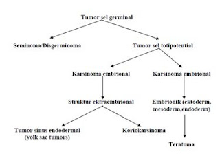
TERATOMA AND DERMOID CYST, DEFINITION, SIGN, SYMPTOMS, ETIOLOGY, DIAGNOSIS AND MANAGEMENT


CHAPTER I
INTRODUCTION

I. 1 OVERVIEW
Teratomas are germ cell tumors are generally composed of multiple cell types derived from one or more of the three germ layers of endoderm, mesoderm and ectoderm. Inconsistent nomenclature is often confused about the various subtypes of teratoma. Teratomas derived from the Greek meaning of the terrace monster, which was first described by Virchow in the first edition of his book published in 1863. Teratoma is divided into three categories: mature teratoma (benign), immature teratoma and teratoma with differentiation monodermal particular degrees depending on the quantity of immature tissue showed the potential for malignancy. Cystic teratomas are generally benign and malignant solid is. Teratomas vary from a benign form of the cystic lesions of well differentiated (mature) to form a solid and malignant (immature). Immature teratomas are malignant germ cell tumor to the three most common after disgerminoma and endodermal sinus tumor. In addition, there also have a specific component (usually squamous) who experienced malignant transformation, but are rarely found.

In 1831, Leblanc created the term dermoid cyst in the literature of veterinary medicine when he succeeded in removing skin lesions at the base of the skull resembles a horse. Both teratomas and dermoid, a term still used and are often used interchangeably. Implications of these elements behawa beginning to resemble the skin with a complementary structure composed of dermoid, whereas teratoma has no such limit. Dermoid now known as the trigeminal and contain various types of tissue.

Monday 9 April 2012

ONCOLOGY / ORTHOPAEDI SURGERY SERIE : SOFT TISSUE CANCER / MALIGNANT FIBROUS HISTIOCYTOMA ( definition, signs, symptoms, diagnosis and management)

INTRODUCTION

In America, more than 2000 people are diagnosed with this tumor each year, mostly one type of cancer suffered by children and adolescents, but few cases are found in adults.
      Cancer around the bone itself consists of several types, namely osteosarcoma, Ewing's sarcoma, chondrosarcoma, malignant fibrous histiocytoma, fibrosarcoma, and Chordoma. Malignant fibrous histiocytoma (MFH) is a soft tissue sarcomas of the most frequently in older adulthood. There are several variants of the MFH, storiform-pleomorphic subtype, myxoid, inflammatory and giant cell. Infammatory and giant cell is a rare subtype. Most MFH tumors found in the soft tissue in the extremities and body. Giant cell MFH found in older patients.

CHAPTER I
DISCUSSION

I.1 Definition
Malignant fibrous Histiositoma (malignant fibrous histiocytoma) is a sarcoma that usually begins in the soft tissues. This condition usually appears as a large mass that is painful and can lead to fractures due to bone destruction by tumor spread. 
Malignant fibrous histiocytoma was first introduced in 1961 by Kauffman and Stout. They describe as a tumor rich in histiocytes MFH with storiform growth pattern. In 1977, MFH is considered as soft tissue sarcomas of the most common of adult life.

I.2 Epidemiology
Malignant fibrous histiocytoma of soft tissue usually appears in patients is approximately 50 to 70 years although it can occur at any age. Malignant fibrous histiocytoma is very rare in people less than 20 years. More men than in women. 
Malignant fibrous histiocytoma has a 44% recurrence rate and metastasis rate by 42%. Incidence of metastases to regional lymph nodes are found as much as 12%. 

I.3 Histiologi
      Histologist is divided into four subtypes:
A. Storiform-pleomorphic
  Of these, storiform-pleomorphic is the most common type, accounting for up to 70% of cases, see Figure 1.

Figure 1: A histologic specimen shows fibrous histiocytoma classic storiform-pleomorphic malignant. Microscopically, the pattern shows fasikula short storiform spindle cells radiating from a central point which is mixed with giant neoplastic cells in the pleomorphic

2. Myxoid
Figure 2: Example of histological type myxoid MFH. So that the tumor will be marked as a variant of myxoid, myxoid tissue must explain at least half of the tumor

LUKA BAKAR / COMBUSTIO / BURN , KLASIFIKASI , DIAGNOSA DAN MANAJEMENNYA ( SKIN BURN DIAGNOSIS AND MANAGEMENT)


LUKA BAKAR / COMBUSTIO / BURN , KLASIFIKASI , DIAGNOSA DAN MANAJEMENNYA

Pendahuluan
Kejadian luka bakar sering terjadi di kehidupan sehari hari di masyarakat. Mengalami kejadian cedera panas adalah salah satu yang paling merusak fisik dan luka psikologis seseorang dan mengalami penderitaan yang amat sangat . Lebih dari 2 juta luka-luka akibat luka bakar yang memerlukan perhatian medis setiap tahun di Amerika Serikat, dengan 14.000 kematian akibat. Kebakaran di rumah bertanggung jawab atas hanya 5% dari cedera bakar tapi untuk luka bakar 50% dari kematian-paling karena inhalasi asap. Sekitar 75.000 pasien memerlukan rawat inap setiap tahun, dan 25.000 dari mereka tetap dirawat di rumah sakit selama lebih dari  bulan 2 tingkat keparahan dari sakit yang terkait dengan cedera ini.


ONCOLOGY SURGERY SERIES : NECK LYMPHADENOPATHY (NECK MASS OR LYMPH NODE) ALGORITMA AND DESCRIPTION


Of the 800 lymph nodes throughout the body is almost (30% of them) 300 lymph nodes in head and neck are so often either metastasis or spreading the infection appears as sweeling of lymph nodes in head neck.
Diagnosis of a lump in the neck requires knowledge of the structure of the normal anatomy of the neck. M. Sternocleidomastoid should be palpated from the top down to ensure there any enlargement of the lymph vessels in the neck jugular.
It is also necessary to distinguish lymph node lump, lump in the thyroid is located in the midline where the thyroid gland and parotid lump which is located in the parotid area preaurikula to the inferior mandibular arch.
Knowledge of anatomy that needs to be understood to include the anatomy of the neck dissection: platisma m, m. sternokleidomastoideus, m. omohioid, m. digastrikus, m. trapezius, mandibular ramus of n. facial, brachial n, n. phrenicus, n. spinal accessory, n. hipoglosus, torachicus duct.


Figure 6. Graph Algorithms evaluation and management of lumps in the neck

Friday 6 April 2012

PLASTIC / Oncologic Surgery SERIES: THE DISTINGUISHING Bell's palsy with Iatrogenic paralise of the facial nerve (etiology, SIGN-symptoms, DIAGNOSIS AND MANAGEMENT)


PLASTIC / Oncologic Surgery SERIES: THE DISTINGUISHING Bell's palsy PARALISE iatrogenic facial nerve (etiology, SIGN-symptoms, DIAGNOSIS AND MANAGEMENT)


The term Bell's palsy is a peripheral facial nerve paresis of unknown cause (idiopathic) and acute. Many are mixed up between Bell's palsy with peripheral facial nerve paresis other unknown causes.
Bell's palsy is found, usually people with facial paralysis learn from friends or family or at the mirror or brush teeth / rinsing. When the patient realizes that he is paralyzed on her face, then he began to feel fear, shame, low self-esteem, cosmetic and sometimes disturbing soul depressed, especially in women and in patients who have a profession that requires him to appear in public. Often the question arises in his heart, if his face could go back to normal or not.

from wikipedia picture
 from freedictionary.com

SERI SEJARAH : PANGERAN KORNEL , PEMIMPIN SUMEDANG PEMBELA RAKYAT DAN BERANI MENENTANG PENJAJAH DEMI RAKYATNYA.


 Sekitar  300 tahun yang lalu ,tepatnya 12 maret 1811 , gubernur jenderal Hindia Belanda  Herman Wilem Daendels  yang sedang memerintahkan membuat jalan dari Anyer (ujung barat pulau jawa  hingga ujung timur pulau Jawa  yaitu Panarukan (kurang lebih 1000 kilometer) lebih jauh baca di http://herryyudha.com . Saat itu terjadi perlambatan pembuatan jalan di sebuah perbukitan batu (cadas) 9 KM sebelum Kota Sumedang . pengerjaan jalan terhambat karena bukit batu tersebut yang sulit ditembus dan curam. Banyak sudah rakyat Sumedang yang disuruh kerja paksa (pekerja RODI) bagaikan budak belian di tempat tersebut dan mereka banyak yang meninggal dunia karena penyakit dan kelaparan. Melihat keadaan tersebut seorang Pangeran S umedang  Kusumah Dinata tidak tega rakyatnya sengsara. Maka pada tanggal tersebut  sang pangeran, mendatangi Daendels. Daendels menyambut sang pangeran dan menyapa Apa kabar pangeran ? sambil menyodorkan tangan kanannya , tetapi pangeran sumedang tidak segera menyambutnya malah sang pangeran menyodorkan tangan kirinya . Daendels sangat terkejut baru kali ini ada seorang pribumi yang berani menghina dan mempermalukan dia.......tetapi sudah terlambat karena pangeran sumedang adalah ahli hikmah , kekuatan ilmunya diatas rata-rata. Sang pangeran langsung bisa menguasai alam bawah sadar  Daendels sehingga seorang Gubernur Jenderal langsung tekuk lutut dihadapan pangeran Sumedang, serta langsung mengsugesti/memerintah tuan Daendels serta  berkata : tuan Daendels rakyat sumedang  banyak yang mati karena ambisi tuan, membuat jalan itu bagus tetapi kalau tuan ingin berhasil tolong perhatikan pekerjanya. Tolong berikan makanan dan peralatan yang baik. Daendels tidak kuasa menantang mata sang pangeran , dia hanya bisa berucap:  iiiiya pangeran, baaik pangeran. Lalu sang pangeran melanjutkan sugestinya : Dan ingat tuan Daendels kenapa saya menyodorkan tangan kiri untuk salaman dengan tuan, karena tangan kanan saya pegang Keris bila tuan tidak mau menuruti nasehat saya maka sekarang juga keris ini akan merobek tubuh tuan. Sebaliknya bila tuan mau berubah memperlakukan pekerja dengan  baik maka saya akan memdukung
tuan membuat jalan                 .
 Babaiik  pangeran ..... hanya itu yang bisa diucapkan Daendels . seterusnya Daendels bengong dan terdiam begitupun para pengawal dan tentara  kompeni. Setelah beberapa lama  pangeran pergi  baru  Daendels tersadarkan, dan berkata : mana pangeran tadi? Pangeran itu  hebat dan berani. Kalo dia mau jadi penasehat saya akan saya berikan pangkat COLONEL . sejak saat itu sang pangeran dipanggil PANGERAN KORNEL  Sejak  peristiwa  itu Daendels memerintahkan supaya pekerja diperhatikan makanan dan peralatannya malah jumlah pekerja ditambah dari daerah lain, malah pasukan Zeni Belanda juga diturunkan sehingga jalan di cadas Pangeran bisa cepat ditembus dan diselesaikan.
 SEJAK SAAT itu daerah bukit batu serta jurang disebut CADAS PANGERAN.  Kalau anda lewat di ujung barat Cadas Pangeran maka akan melihat tugu ada dua orang yang lagi salaman yang satu pake tangan kiri (Kornel) yang satu lagi pake jubah salaman tangan kanan (Daendels).

Saya sebagai generasi penerus / keturunannya Pangeran Kornel tetap memelihara ilmu ilmu kasumedangan......yang mungkin nanti akan saya tulis di blog ini untuk melestarikan sampai generasi  yang akan datang.
 Sayang peristiwa istimewa ini luput dari para ahli sejarah. Di jawa barat  tidak terjadi perang besar ,saat pembuatan jalan Daendels karena gubernur jendral Daendels bisa dikendalikan dengan bijaksana ( di daerah lain banyak bentrokan / perang ).
Di  jawa barat penduduknya banyak  . luas jawa barat hampir sama dengan luas nedherland. Banyak orang jawa barat yang pintar ,arif serta bijaksana namun penulis perhatikan , dari pemerintahan ke pemerintahan orang jawa barat mulai terpingirkan (mungkin yang lain tidak memperhatikan) sehingga  kepemerintahan di kita sering labil dan mudah digoyang karena orang orang pinter,arif serta bijaksana yang notabene banyak di jawa barat tidak diikutsertakan secara optimal  dan proposional.
Dilain pihak juga banyak  orang jawa barat  yang sudah jadi pemimpin ,baik level lokal maupun nasional tidak ingat lagi pada rakyat kecil,  mereka sudah jadi egoistis, melupakan budaya asal  dan kemaruk , hanya mementingkan diri sendiri dan kelompoknya.
Kalau ingat sejarah  Majapahit juga sulit  menguasai Jawa Barat sampai terjadi peristiwa rekayasa di Bubat kisah putri Pitaloka. mangkanya di jawa barat tidak pernah ada daerah/jalan Majapahit,Gajah mada ataupun Hayam wuruk.  Sejarah juga mencatat Sultan Agung bisa menggempur batavia karena di bantu orang jawa barat,,,,,,,,kalau mau maju Indonesia jangan lupakan orang jawa barat yang baik,bukan orang jawa barat yang buruk/busuk .chauvinisme ? oh no, tentu saja tidak. bukankah segala sesuatu harus dimaximalkan dioptimalkan ,secara seimbang serta proposional dan profesional.    ini anjuran ..... percayalah padaku . mudah mudahan pemerintahan mendatang kedepan bisa memahami ini kalau mau stabil.

Thursday 5 April 2012

SIGN, SYMPTOMS, DIAGNOSIS, AND MANAGEMENT SPINA BIFIDA


CHAPTER I
INTRODUCTION

The Spina bifida is a congenital defect of the posterior arch of the spine due to failure of closure of the neural elements of the spinal canal in the early development of embryos. (1,2,3)
In the early stages of neural plate formation of a gap is formed which then form the neural neural tube. Pipe is then a neural network of the brain and spinal cord. While in the womb, the tissue that forms neural tube does not close or not closed properly. This led to the opening in the vertebrae, which surrounds and protects the spinal cord. The process of neural tube closure takes place during the fourth week of embryonic life, and usually before a woman knows her pregnancy and ended. Neuralisasi process began at the dorsal midline and continues to sefal and caudal direction. The most recent closure occurred at the posterior end of the day-to-28. (3.4)
Sometimes it does not close the neural groove, this is because the error induced by chorda spinalis which lies beneath or under the influence of environmental teratogenic factors neuroepitel cells. Neural networks in this case remains open to the outside world. Disruption of this process causes neural tube defects which are then classified as disrafisme. Disrafisme divided into two cranial and spinal. (3,4)
Disrafisme spinal / myelodysplasia is a congenital anomaly of the spinal fusion caused by the failure of the structures at the midline. When the lesions confined to the bones (arch) whether one or more posterior levels, the disorder is referred to as spina bifida. (4, 5, 6)
If the neural elements involved it will cause paralysis and loss of sensation and sphincter disorders. The degree and localization of defects that occur vary. Light on the circumstances which may be found only a failure of fusion of one or more of the posterior arch of the vertebra at the lumbosacral region. Sometimes this disorder does not cause significant clinical symptoms. (1, 2,4,7)
Often the event of a defect in the posterior arch will give rise to disorders of the skin surface of the cover, which looks like a dimple, tuft of hair, skin or sinus fat mass.
Spina bifida can be classified into two types namely, spina bifida occult spina bifida and aperta (cystica). (1)


CHAPTER II
REVIEW REFERENCES

DEFINITION 
Spina bifida is a developmental anomaly characterized by defective closure of the bones in the spinal cord sheath so that the lining of the spinal cord and meninges may protrude (spina bifida cystica), or does not protrude (spina bifida occulta) .(2)
Several hypotheses of spina bifida include: (4)
1. Cessation of neural tube formation process as a particular cause
2. Excess pressure in the central canal of the newly formed rupture surface, causing neural tube
3. The damage to the walls of the newly formed neural tube as a cause.

Protrusion of the spinal cord and meningens cause damage to the spinal cord and nerves, resulting in decreased or impaired function of the body parts supplied by these nerves, or at the bottom. Symptoms depend on the anatomical location of spina bifida. Most occur in the lower back, the lumbar or sacral region, due to the closure of the vertebrae in place at the end of this section.
         

                                                                 Figure 1. Spina Bifida

Wednesday 4 April 2012

TYPE OF NEUROFIBROMATOSIS OR VON RECKLINGHAUSEN'S DISEASE ( BENIGN DISORDER BUT CREEPY DISEASE ) : DEFINITION, ETIOLOGY,SIGN , SYMPTONS ,PROGNOSiS, DIAGNOSIS AND MANAGEMENT


CHAPTER I
INTRODUCTION

Neurofibromatosis is an autosomal dominant disorder that affects the bone, nervous system, soft tissue, and skin. At least 8 different clinical phenotypes of neurofibromatosis have been identified that are associated with at least two genetic disorders. Clinical manifestations increase over time. Neurological and developmental problems toward malignancy might come afterwards.

Neurofibromatosis type 1 have varying phenotypic expression including dermatologic manifestations. Some patients may have a particular expression of the skin, while others may have life-threatening complications.

A neurocutaneous condition, neurofibromatosis may involve almost any organ system. Thus, the signs and symptoms can vary widely demonstrated. Two major subtypes exist: neurofibromatosis type 1, also known as von Recklinghausen neurofibromatosis, which is the most common subtype and is referred to as the neurofibromatosis, and neurofibromatosis type 2, the so-called central neurofibromatosis is a genetic disorder associated with bilateral vestibular schwannomas multisystem, schwannomas spinal cord, meningiomas, gliomas, and juvenile cataracts, skin with lack of features.


CHAPTER II
REVIEW REFERENCES

II. A. DEFINITION
Neurofibromatosis is a genetic disorder that interferes with the growth of cells in the nervous system, causing tumors to form in nerve tissue. These tumors can occur anywhere in the nervous system, including the brain, spinal cord, and nerves of large and small. Neurofibromatosis is usually diagnosed in childhood or early adulthood.

Tuesday 3 April 2012

MANFAAT TUMOR MARKER UNTUK DETEKSI DINI, DIAGNOSA,RESPON TERAPI DAN PROGNOSA PENYAKIT KEGANASAN


BAB I

PENDAHULUAN
Kemajuan ilmu pengetahuan dan teknologi dalam hal imunologi, kemajuan membawa bidang diteksi dini onkologi (kanker). Juga termasuk dalam diagnosis masalah, menentukan prognosis dan pemantauan kanker baru-baru ini banyak perhatian diarahkan pada berbagai zat yang seharusnya memberikan petunjuk tentang perkembangan tumor ganas dan komplikasi itu causes.Identification substansi diharapkan untuk membantu mendirikan diagnosis, menentukan prognosis dan memprediksi perjalanan penyakit.
Dengan perkembangan teknologi laboratorium, terutama perkembangan di bidang bioteknologi, sekarang mungkin untuk mendeteksi penanda ganas, tidak hanya dalam lingkungan ekstraselular atau pada tingkat sel tetapi juga pada tingkat molekuler sehingga penanda untuk ganas tidak hanya digunakan untuk atas tujuan tetapi, penanda molekuler tertentu ganas, juga digunakan untuk mendeteksi sel kanker sisa (penyakit sisa minimal, MRD), bahkan dalam keadaan tertentu dapat digunakan sebagai faktor prediksi keganasan atau faktor risiko.

Karena gejala klinis kanker diawali dengan pertumbuhan sel yang tidak terkendali, kanker sebagai penyakit yang disebut sel atau jaringan patologis dan penyakit organ karena sel-sel yang tumbuh di luar kendali dapat menyusup ke jaringan organ dan mengganggu fungsi organ yang bersangkutan. Tetapi sekarang diterima secara luas bahwa kanker disebabkan oleh akumulasi kelainan atau mutasi gen tertentu, karena kanker disebut juga penyakit genetik. Selain menyebabkan gejala yang secara langsung disebabkan oleh kanker atau anak-anak sebarnya, kanker dapat memberikan efek sistemik lainnya yang timbul sebagai respon tubuh terhadap kanker. Gejala yang dikenal sebagai sindrom yang sering menyebabkan komplikasi paraneoplasia atau penderitaan yang lebih buruk, karena kanker juga dapat dianggap sebagai penyakit sistemik.
Dengan demikian, dalam menggunakan dan menafsirkan hasil penanda tumor dan lain tes laboratorium klinis untuk manajemen kanker perlu dipahami kelainan genetik yang sama sekali berbeda, sel dan organ serta respon tubuh terhadap kanker.
Secara umum, penanda tumor adalah perubahan yang dapat dideteksi dan menunjukkan adanya tumor, tumor terutama ganas atau kanker.

Monday 2 April 2012

SERIE BEDAH DIGESTIVE: ILEUS OBSTRUKTIVE DAN PARALITIK ,PERITONITIS SERTA PERFORASI USUS DALAM GAMBARAN RADIOLOGI


KATA PENGANTAR
Dalam praktek sehari-hari di bag bedah, terutama ketika kasus darurat, kita sering menemukan kasus ileus gastrointestinal di mana kadang-kadang kita harus hati-hati membedakan ileus paralitik dengan ileus obstruktif . Berikut akan disajikan pada gambar radiologis untuk memahami gangguan atau kasus ileus untuk memfasilitasi dan mendukung diagnosis sehingga pengobatan lebih cepat dan akurat, tentu saja, gambaran radiologis bukan dukungan hanya untuk diagnosis. Penampilan klinis adalah petunjuk untuk dasar diagnosis  kita melakukan tindakan.


Ileus: berlalunya isi usus yang interupsi adalah tanda obstruksi usus akut yang membutuhkan bantuan segera atau tindakan. Obstruktif ileus: kerusakan atau kehilangan bagian dari isi usus yang disebabkan oleh obstruksi mekanis. Ileus obstruktif = Obstruksi

Ileus paralitik: kerusakan atau kehilangan bagian dari isi usus yang disebabkan oleh selain penyumbatan mekanis. Ileus paralitik ileus adinamik = non-obstruktif ileus =
Lokasi obstruksi:
Lokasi yang tinggi: duodenum untuk jejunum
Rendah letak: usus besar - sigmoid - rectum
iliocecal terbatas oleh persimpangan

Stadion:
Parsial: sebagian menyumbat lumen
Sederhana / Lengkap: menyumbat lumen seluruh
Pencekikan: sederhana dengan vasa tercekik


Style Energy Efficient, practical,economical, and healthy !!

Style Energy Efficient, Practical,Economical, and Healthy!
        We often think that energy-efficient and more difficult and complicated to do, think think again. A lot of really easy and simple way we can do every day.

      Listen here, simple steps you can do to conserve energy use at home.

  •  Make a habit of walking or bicycling when traveling close enough.
  •  We have to get used to clean the air filter located in a variety of household items, such as air conditioners and vacuum cleaners, every two or three months. Dirty air ducts will further increase the energy use for the equipment is turned on.
  •  We need to make sure every time you close the refrigerator door closed. Open the refrigerator door will increasingly absorb the energy use for refrigerators need more energy to keep working.
  •  If we're going to use a washing machine, make sure the tubes are used capacity is full then we began to wash. Tube containing a few clothes require the same energy with a full tube. For traditional drying rather than tumble drying on a washing machine because it is much more energy efficient.
  • How to Remove the plugs always plugs-we are not using, including cell phone charger. Leaving the cord still attached in a state, it spends energy. In order to simplify, we can use the plug with a colored button to stay pressed to stop the flow of electrical energy to the cable.
  • Must Be sure you use the sleep mode or hibernate feature on your computer. So, when not in use, no electrical energy is wasted. We can save energy up to 15 percent by doing this.
  • Try to Replace all regular light bulbs with energy-efficient CFL. They use two-thirds the energy of traditional bulbs and last up to 10 times longer.
  • We do not forget to use the lid when you're boiling food. Cover the pan very helpful wasting a lot of heat energy is wasted.
  •  Use a detergent as needed. The use of plastic bag/ wrap as necessary.