CHAPTER I
INTRODUCTION
It is a benign neoplasm that is locally infiltrate and possibly malignant (10-15%). Used to be called Cystosarcoma phyllodes are benign tumors that occur almost exclusively on women's breasts. Its name comes from the Greek word sarcoma, which means the fleshy tumor, and phyllo, meaning leaf. So called because the tumors display characteristics of a large, malignant sarcomas, such as display-leaves when cut, and the epithelium, cyst-like space when viewed histologically. Because most of the tumor was benign tumor is also called filoides.
Although metastatic tumors are not benign, but they have a tendency to grow locally aggressive and recurrent. Similar to sarcoma, malignant tumor hematogenous metastasis. Filoides tumor pathological picture is not always predict the clinical behavior of neoplasms, hence in some cases there is uncertainty about the classification level of the lesion.
The characteristics of malignant tumor filoides are as follows:
· Recurrent malignant tumors are more aggressive look than the original tumor.
· Pulmonary metastasis is the place most frequently, followed by bone, heart, and liver.
· The symptoms for metastatic involvement can occur immediately, until a few months and no later than 12 years after initial therapy.
· Most patients with metastases died within 3 years of initial therapy.
· There is no treatment for systemic metastases occurred.
· Roughly 30% of patients with malignant tumors filoides died of the disease.
Filoides tumor etiology is not known, it is estimated associated with fibroadenoma in some cases, because patients may possibly have a second histological lesions in the same tumor. However, if the tumor develops filoides of developing fibroadenoma or both together, or whether the tumor can arise de novo filoides, is still unclear
This tumor has a smooth texture, sharply bounded and usually move freely. This tumor is relatively large tumor, with an average size of 5 cm. However, lesions> 30 cm have been reported.
Haagensen reported roughly one for every 40 filodes tumor fibroadenoma. Distribution of age according to Haagensen majority occur between the ages of 35 and 55 years.Bilateral tumors are very rare and rarely also occur in patients under age 20, first appeared benign react.
Mammography and ultrasound examination (Figure II.03) are generally important in the diagnosis of breast lesions, but they are notoriously unreliable in differentiating benign tumor from filoides malignant form of the condition or of fibroadenoma. Thus, the findings on imaging studies is not a definite diagnosis. Open excision biopsy of breast lesions smaller or incisional biopsy for larger lesions is a surefire method for diagnosing tumor filoides.

image 03. Mammography image of the tumor filoides
In most cases tumor filoides, performed wide excision. With boundary eksisinya namely: limit of 2 cm for small tumors (<5 cm) and a limit of 5 cm for large tumors (> 5 cm) has been recommended. Another consideration in the surgical treatment are:
· If the ratio of tumor to normal breast tissue is high enough, to give a satisfactory cosmetic result is a segmental excision. Total mastectomy with or without reconstruction is an alternative option.
· A more radical procedure is not generally justified.
· Conduct an axillary lymph node dissection only for clinically suspected nodes are malignant. However, virtually all the nodes are reactive and do not contain malignant cells.
Is a rare variant of the fibroadenoma. Cystosarcoma phyllodes are responsible for less than 1% of all benign and malignant breast lesions. His name is wrong because it is rarely malignant and are usually not cystic. Their origin from the existing cellular fibroadenoma which now contains one or more components of mesenchymal origin
These lesions are less frequent compared to FAM. Growth pattern of these lesions tend to expand rapidly and surrounding area. Although not all, there are some forms that cystic2 lesion is often also called cystosarcoma phyllodes. These lesions are relatively benign and rarely bermestasis and pathological mitosis, but 15% can be malignant and hematogenous spread (high grade). Therefore kabanyakan local lesions, then it can be cured by local excision, malignant lesions tend to grow back, but it also tends lokal.Tumor have at any age, but most are around the age of 45 years.
CHAPTER II
TUMOR PHYLLOIDES
Anatomy and Physiology
A. ANATOMY
To be able to recognize breast cancer journey well and understand the basics of surgery in breast cancer it is important to know the anatomy of the breast itself.
Breast lies in hemithoraks right and left with the following limits:
A. Boundaries of the breast that looks from the outside:
- Superior: ribs II or III
- Inferior: VI or VII ribs
- Medial: the edge of the sternum
- Lateral: anterior axillary line.
2. The boundaries of the real breast:
- Superior: almost to the clavicle
- Medial: midline
- Lateral: m.latissimus dorsi
Structure of the Breast
Breast consists of various structures:
- Parenchymal epithelial
- Fat, blood vessels, nerves and lymphatic channels
- Muscle and fascia
Figure 1. Anatomy of the breast.
Parenchymal epithelial formed by approximately 15-20 lobes, each of which has its own channel to drain the product, and comes down to putting susu.Tiap lobe-lobule lobules formed by each of which consists of 10-100 asini grup.Lobulus-lobule This is the basic structure of the mammary gland.
Breast is covered by the superficial pectoral fascia of the anterior and posterior surfaces which are connected by a ligament which serves as a buffer Cooper.

Breast Lymphatic System
Lymph vessels
A. Axillary lymph vessels:
This axillary lymph vessel draining lymph from the area surrounding the areola mammae, the lateral quadrant and the upper lateral quadrant of the breast.
2. The internal mammary lymph vessels:
These lymph channels drain lymph from the inside and the medial breast. This vessel runs above the pectoralis fascia and through the fascia, and into major m.pektoralis. Then the road to the medial together with penetrating perforating system m.interkostalis and empties into the internal mammary lymph nodes. Of the internal mammary glands, lymph flow through the internal mammary lymph trunks. Some will lead to v.cava, some will lead to the duct torasikus (to the left) and lymph duct artery (to the right side).
3. Lymph vessels in the medial edge of the lower medial quadrant of the breast. These vessels take a walk along the superior epigastric vasa, through the rectus fascia and into m.rektus abdominis. This channel empties into the lymph nodes that terletek preperikardial anterior edge of the falciform ligament above the diaphragm. Lymph nodes also contain lymph from the diaphragm, falciform ligament and the antero-superior hepar.Dari this gland, lymph flow through the internal mammary lymph trunks.
Lymph glands
- Axillary lymph nodes
There are six axillary lymph node group:
External mammary lymph nodes. Strands gland is located below the lateral edge m.pektoralis mayor, along the medial edge of the axilla. This group is divided into two groups.
a) The superior: The lymph nodes are located as high as intercostal II-III.
b) Group inferior.Kelompok lymph nodes are located as high as intercostal IV-V-VI.
Lymph nodes of the scapula.
Lymph nodes are located along the vasa-torako subscapular and dorsalis, ranging from a vein subscapular v.aksilaris branching, up to their entrance into the dorsal v.torako-m.latissimus dorsi.
3. Central lymph node (Central Nodes)
Lymph nodes are located in fatty tissue in the center of the armpit. Sometimes some of them are located very superfisisal, under the skin and fascia at the center of the armpit, at approximately the mid-axillary fold front and back. Lymph nodes are glands that are relatively easily palpable. And the axillary glands of the largest and most in number.
4. Interpektoral lymph nodes (Rotter's Nodes)
Lymph nodes are located between m.pektoralis

Figure 2. Breast lymphatic system.
5. V.aksilaris lymph nodes.
These glands are located along the lateral v.aksilaris, ranging from white m.latissimus dorsi tendon to the medial bit of branching v.aksilaris - v.torako-acromion.
6. Subklavikula lymph nodes.
These glands are located throughout v.aksilaris, ranging from slightly medial branching v.aksilaris - v.torako-acromion to where v. axillary disappeared under m.subklavius tendon. Axillary lymph glands is the highest and termedial located. All lymph nodes derived from the glands into the axillary lymph glands. The entire axillary lymph nodes are located under the fascia kostokorakoid.
7. Lymph nodes prepektoral
Sometimes located under the skin or in the upper lateral quadrant of the breast tissue called fascia prepektoral because it is located above the pectoral.
8. The internal mammary lymph nodes.
These glands are spread along the internal mammary lymph trunks, approximately 3 cm from the edge of the sternum. Located in the fat over the fascia endotorasika, in between the ribs. Estimated number of these glands have 6-8 pieces.
2. PHYSIOLOGY
Phase of breast development arising as a result of the effects of ovarian hormone secretion mamotropik and anterior pituitary, where the breast had three kinds of changes that affected the hormone:
A. The first change is the start of the lifetime of the child through puberty, a period of fertilization, to klimakterium and menopause. Since puberty the hormones estrogen and progesterone production in the ovaries and pituitary hormones has led to the emergence of the ducts and acini grown.
2. The second change is the change in accordance with the menstrual cycle, about day-to-8 menstrual breasts become larger and in a few days before the next menstrual period maximal enlargement. Sometimes - sometimes uneven bumps arise. For several days before menstruation breast pain became so tense and physical examination, especially palpation is not possible. At that time the examination is not useful because the mammogram image contrast is too large glands. Once menstruation begins, everything is reduced.
3. Changes that occur during pregnancy and lactation. In pregnancy the breasts become huge because lobul duct epithelium and alveolar ducts proliferate and grow a new duct.Secretion of prolactin from the anterior pituitary hormones trigger lactation. The milk produced by the cells of the alveoli, filling the acini, and then expelled through the ducts to the nipple.
Breasts are composed of fatty tissue containing glands responsible for milk production during pregnancy and after childbirth. Each breast is made up of about 15-25 lobes in groups, called lobules, mammary gland, and a formation like sacs that contain milk (alveoli). Channel to drain the milk to the nipple are called ducts. About 15-20 channels will go dark circular part around the nipple (areola) forming part of the store breast milk (ampullae) before exiting to the surface.
Both breasts do not always have the same size and shape. Of the breasts begin to form a complete one or two years after menstruation pertamakali.Hamil and breast feeding will lead to increase in size and will experience a diminution (atrophy) after menopause.
Breasts will cover most of the chest wall. Breast is limited by the collarbone (clavicle) and the breastbone (sternum). Breast tissue could reach into the underarm area and the muscles that are on the lower back until your upper arms (latissimus dorsi).
Lymph nodes consist of white blood cells that are useful to fight the disease. Drained by the lymph nodes of breast tissue through lymph channels and into the lymph glands around the breast till the armpit and collarbone. Lymph nodules play an important role in the spread of breast cancer especially in the armpit lymph nodules.
DEFINITION
Cystosarcoma phyllodes derived from the Greek word, sarcoma, which means the fleshy tumor, and phyllo, meaning leaf. These tumors display characteristics of a large, malignant sarcomas, such as take-leaf appearance when cut, and displays the epithelium, cyst-like space when viewed histologically. Because most of the tumor was benign, his name can be misleading. Thus, the preferred terminology is now filodes tumor.
Johann Muller is the man who first gave the name 'cystosarcoma phyllodes' in 1838, because these tumors are often cystic and classically has a projection like a leaf into it.
These tumors are usually large and growing very rapidly. These tumors may be benign or malignant and can spread to other parts of the body. Also called CSP (Cystosarcoma phyllodes) or tumor filodes. Is a type of connective tissue neoplasm arising from intralobular breast stroma.
Synonym
Cystosarcoma phylloides, cystosarcoma phyllodes, filodes tumor.
FREQUENCY
There was no difference in tumor frequency filodes among patients in the United States and patients in other countries. Tumor filodes estimated at around 1% of total breast neoplasms.
v Mortality / Morbidity
Due to limited data, the percentage of malignant than benign tumors filodes terdefenisi not well. The reports indicate that approximately 80-95% of tumors are benign and that filodes about 10-15% are malignant.
Although metastatic tumors are not benign, but they have a tendency to grow locally aggressive and recurrent. Similar to sarcoma, malignant tumor hematogenous metastasis. Unfortunately, the picture is not always pathological tumor filodes predict the clinical behavior of neoplasms; therefore in some cases there is uncertainty about the classification level of the lesion. The characteristics of malignant tumor filodes are as follows:
Recurrent malignant tumors appear more aggressive than the original tumor
Pulmonary metastases is a place most often, followed by bone, heart, and liver
Symptoms for metastatic involvement can arise from as soon as a few months to at least 12 years after initial therapy
Most patients with metastases died within 3 years of initial therapy
There is no treatment for systemic metastases occurred
Roughly 30% of patients with malignant tumors filodes died of the disease
v RAS, SEX AND AGE
· Predilection for the tumor seems to be no filodes.
· Tumor filodes muncur almost exclusively in women. Rare case reports have been described in men.
· Tumor filodes can occur at any age; but middle age is the fifth decade of life.
· Some juvenile fibroadenomas in adolescents can look like a tumor histologically filodes; however, they behave like benign fibroadenoma other.
Etiology
Tumor filodes significantly associated with fibroadenoma in some cases, because patients can have both histological lesions and the lesions may be seen in the same tumor. However, if the tumor develops filodes of developing fibroadenoma or both together, or whether the tumor can arise de novo filodes, it is not clear. Noguchi and colleagues have studied this question by clonal analysis in three cases of fibroadenoma and tumor filodes obtained sequentially from the same patient. In each case, the tumor showed a monoclonal and inactive alleles of the same. They argue convincingly that the tumor has the same origin filodes with fibroadenoma, fibroadenoma may develop into a tumor specific filodes.
Interesting study by Yamashita et al, observed immunoreactive endothelin 1 (IRET-1), examples of behavior which modern science to explain the mechanism that will surely prove important in understanding both normal function and pathology of the breast, while allowing a shift in emphasis from model to study human rodentia . IRET-1 tissue levels measured in extracts from four tumor filodes and 14 fibroadenomas. Immunoreactive endothelin 1 can be proved in all cases, but their levels were significantly higher in tumors than in fibroadenomas filodes. Endothelin 1 in principle is a powerful vasoconstrictor, but also has many other functions. He has caused a modest stimulation of breast fibroblast DNA, but can be combined with insulin-like growth factor 1 (IGF-1) to create a strong stimulation. ET-1 is absent in normal breast epithelial cells, but ET-1-specific receptor found on the surface of normal stromal cells. ET-1 receptors found on the cell surface of tumor stromal cells filodes but immunoreactive cells found in epithelial cells but not stromal cells, suggesting that ET-1 synthesized by epithelial tumor cells filodes. Thus it provides the possibility of a paracrine mechanism to stimulate rapid growth of the stroma is always seen with a tumor filodes.
What is important is that the tumor should not be confused with filodes pure sarcoma (without epithelial elements at all), to have a greater degree of malignancy and lump the two together could obscure the most fundamental properties of benign tumor filodes.Imunositokemistri and electron microscopy showed that the stromal cells in both benign and malignant tumors filodes is a mixture of fibroblasts and miofibroblas. These techniques are freed from leiomiosarkoma and mioepitelioma differences, which may mimic tumor filodes show a completely different reaction.
Pathophysiology
Filodes tumor is non-epithelial neoplasms of the breast are the most common, although it only represents 1% of breast tumors. This tumor has a smooth texture, sharply bounded and usually move freely. This tumor is relatively large tumor, with an average size of 5 cm.However, lesions> 30 cm have been reported.
TUMOR BEHAVIOR
While the trend clearly shows filoides tumor to recur locally if excised with close limits, local or distant metastasis is rare. In fact, tumors are considered as benign after thorough histological studies can be expected to have a good prognosis, especially if initially treated with complete excision. Tumors are histologically malignant (sarcoma filoides) unpredictable behavior.
Single-center study of 32 cases give a reasonable indication of the behavior. Benign tumors do not show recurrence if excised completely, but half (6 of 13) who had complete excision of non-local recurrence. There is no visible recurrence after complete excision of the four boundaries and four malignant tumors, but incomplete excision of malignant tumor diseases of the chest wall leads to non-controlled.
In general, the local recurrence of benign tumors remain benign, but malignant transformation to occur and the explosion of malignancies have been reported after 15 episodes of local recurrence of benign.
Prognosis good overall look at the series of Haagensen, where only four of 84 patients known to have metastases. While we found a local recurrence in patients, none of which had metastasis. Latest series of 66 cases from the Mayo Clinic confirmed that most low-degree-behaved, non-metastatic tumors, but both histological evaluation and DNA analysis by flow cytometry provides a reliable assessment of the behavior of the individual tumor.
CLINICAL
Haagensen reported roughly one for every 40 filodes tumor fibroadenoma. Broad distribution of ages, from 10-90 on the Haagensen series of 84 patients, but with the majority between 35 and 55 years. Bilateral tumors are very rare, although an exceptional case of three separate tumor in bilateral axillary ectopic breast tissue is normal breast also been reported. Filodes rare tumor in patients under age 20, when it appeared to react primarily with benign way, without regard to the histological pattern. Also been described in similar mammary gland in the vulva, breasts, and men in the prostate and seminal vesicles.
Most tumors grow rapidly into a large size before the patient arrives, but tumors are not settled within the meaning of the carcinoma. This is because they are not particularly invasive; size of the tumor may occupy most of the breast, or entirely, and cause pressure ulcers on the skin, but still showed some mobility in the chest wall.
Anamnesa
Patients with a breast mass in particular appear hard, moving, clear boundary, not soft
A small mass can quickly grow in size in a few weeks before the patient seeks medical attention
Rare tumors involving the nipple-areola complex, or to skin ulceration
Patients with metastases can occur with symptoms such as dyspnoea, fatigue, and bone pain
Physical examination
Realize the hard breast mass, moving, boundary-clearly, not soft


Figure 3. Breast Examination
Is odd, cystosarcoma phylloides tend to involve the left breast more often than the right breast
Appearance on the skin may look slick and translucent enough to reveal the underlying breast veins
Physical findings (eg, a moving mass with clear borders) is similar to that of the fibroadenoma
Filoides tumors commonly manifest as a greater mass and showed rapid growth
Mammography findings (eg, circular display with smooth boundary density) is also similar to embedded fibroadenoma
Recurrent Malignant tumors are more aggressive look than the original tumor
Pulmonary metastases is a place most often, followed by bone, heart and liver
Symptoms for metastatic involvement can arise from as soon as a few months to at least 12 years after initial therapy
Most patients with metastases died within 3 years of initial therapy
There is no treatment for systemic metastases occurred
Count of roughly 30% of patients with malignant tumors filoides died of the disease
Examination support
· The laboratory
No hematologic tumor markers or other blood tests that can be used to diagnose cystosarcoma
· Imaging Studies
On a mammogram, Phyllodes tumor will have a clear boundary edges. Both mammogram or breast ultrasound can distinguish clearly between fibroadenoma and Phyllodes tumor is benign or malignant. Types of breast tumors are usually found near the microcalcifications.The cells from the biopsy needle can be tested in the laboratory but rarely give a clear diagnosis, because these cells may mimic carcinoma and fibroadenoma. On surgical biopsy tissue sections that will produce will provide a better cell sample and will result in proper diagnosis for a Phyllodes tumor.

Figure 4. Mammography image cystosarcoma
Breast MRI can assist in the surgical removal of phyllodes tumor tissue. A study in Italy comparing mammograms, breast ultrasound and MRI of Phyllodes tumor reported that MRI gives the most accurate and it helps the surgeon plan the tumor in running their operations. Even if the tumor is close enough to the muscles of the chest wall, breast MRI can provide a better picture of phyllodes tumors than a mammogram or ultrasound.
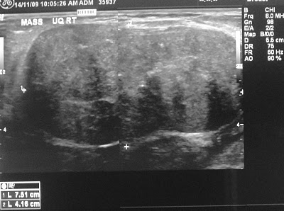
Figure 5. Ultrasound picture

Procedure
FNA for cytology is usually not sufficient for tumor diagnosis filoides. Needle biopsy is more reliable, but still there are sampling errors and the difficulty in distinguishing the lesion from a fibroadenoma
Open excision biopsy of breast lesions smaller or incisional biopsy for larger lesions is a surefire method for diagnosing tumor filoides
Histologic findings
All filoides tumor stroma contains components that can be varied in histologic appearance of the lesions to other lesions. Generally, benign tumors showed filoides striking increase in the number of regular fusiform fibroblasts in the stroma.

Occasionally, highly anaplastic cells with miksoid changes were observed. Cellular Atipia high level, with increased selularitas stroma and increased number of mitosis, is almost always observed in the malignant form of cystosarcoma phylloides. In ultra-structural, in the form of benign tumors and malignant filoides, nucleoli may reveal a mesh nukleolonema sisterna coarse and abundant in the endoplasmic reticulum.

Differential Diagnosis
Angiosarcoma
Breast cancer (Cancer)
Another issue to consider:
· Juvenile fibroadenoma
· Giant fibroadenoma
· Inflammatory carcinoma
· Sclerosing adenosis
· Radial scar
· Fat necrosis
· Fibrocystic Changes
· Breast abscess
· Adenocarcinoma
· Mastitis
MANAGEMENT
Age is important in the management of these lesions. Under the age of 20, it should be treated with enucleation, because they almost always behave in a benign manner.
Aspiration cytology can suggest the diagnosis of tumor histology filoides but more precise in the core needle biopsy is needed before planning treatment.
Another situation in older patients. Some surgeons have enough experience to be dogmatic about its management. Haagensen reported one of the largest series, and recommend a wide local excision as the primary approach to the treatment of benign tumor filoides. He had a local recurrence rate of 28% among 43 patients treated with local excision, with follow-up of at least 10 years. However, only 3 of these recurrences requiring secondary mastectomy, and none of them died of this tumor. Only 1 of 21 patients treated with mastectomy (simple or radical) had local recurrence; this is a rapidly filoides sarcoma causing local and systemic metastasis. Higher recurrence rate for benign than malignant tumors filoides have been reported in several series, reflecting a more simple surgical approach to tumors estimated to be less serious.
It is clear that no-complete excision is the major determinant of recurrence in benign lesions and secondary schools. Why the high recurrence was reported from most of the series while it is so well shown? There are two main reasons: failure to anticipate the possibility of failure to define tumor filoides and refinement that will assure complete excision. The former can be found only with high level of suspicion, and a triple assessment at all masses prior to surgery. Particularly important to avoid an excision biopsy as a diagnostic procedure because it is almost impossible strongly influence the extent of excision biopsy cavity, where it is performed as primary procedures while the tumor remains in situ. For this reason, histological diagnosis to be made by needle-core biopsy, or at least no more procedures than incisional biopsy.
Macroscopic complete excision, with the proposed limit of 1 cm, can be ensured by proper technique. With normal excision technique while placing traction on the masses, easy to perform dissection too close to the tumor at some point dissection. Reliable way to avoid this is to a surgeon place the fingers of the left on the mass, and cut out the finger, with traction only on the surrounding breast tissue.
For small lesions where the diagnosis is suggested by the triple assessment or macroscopic appearance (soft, brown, fleshy appearance), the tumor should be excised with a 1-cm border of normal breast tissue. If histologinya benign, it is sufficient management, with excision quadrantic (quarter-circle) for intermediate lesions. Where the diagnosis was first identified on histologic examination of excisional biopsy specimen, excision of scar tissue quadrantic recommended in order to ensure that qualified local clearance.
For large lesions and recurrent lesions, a good cleaning would involve a near-total mastectomy, and we preferred a simple mastectomy, with reconstruction of the medium should be expected of patients. There is some evidence of increasing incidence of breast carcinoma associated, simultaneously or subsequently, in patients with tumor filoides and this is an additional reason for the long-term follow-up to careful examination of such patients.
Surgical therapy
In most cases of cystosarcoma phylloides, do the normal wide excision, with a circle of normal tissue. There are no rules about the size limit. However, 2 cm limit for small tumors (<5 cm) and a limit of 5 cm for large tumors (> 5 cm) has been recommended.
Lesions should not be "shelled out", as you might do with fibroadenoma, or without an acceptable recurrence rate increases.
If the tumor to breast ratio is high enough to do segmental excision, total mastectomy, with or without reconstruction, is an alternative.
A more radical procedure is not generally justified.
Axillary lymph node dissection do only to nodes that are clinically suspected. However, virtually all the nodes are reactive and do not contain malignant cells.
COMPLICATIONS
Like most breast surgery, postoperative complications of surgical treatment of tumors filoides include the following:
Infection
Seroma formation
Local recurrence and / or distant
Prognosis
Although cystosarcoma phylloides regarded as clinically benign tumor, the possibility of local recurrence after excision is always there, especially with lesions showed malignant histology. Tumor after initial treatment with wide local excision, which locally recurrent ideally treated with total mastectomy.
Metastatic disease was observed particularly in the lungs, mediastinum and bones.
Serving a variety of clinical
If a tumor is benign, long-term prognosis was good following adequate local excision
If the local recurrent tumor after excision recara, we then performed a local excision or total mastectomy, especially curative
REFERENCES
- Ramli muchlis. KUMPULAN KULIAH ILMU BEDAH.1995.Jakarta : Binarupa aksara.Halaman 355
- Jong de wim. BUKU AJAR ILMU BEDAH EDISI 2.2004. Jakarta : EGC. Halaman 391-393
- Manning. MAJOR DIAGNOSIS FISIK EDISI IX. 1996. Jakarta : EGC. Halaman 366
- Schwartz. INTISARI PRINSIP-PRINSIP ILMU BEDAH EDISI 6. 2000. Jakarta : EGC. Halaman 233
- muel-muel.blogspot.com/.../neoplasma-jinak-payudara.htm
- www.bidadariku.com/idpayudara2.php?kode=90
- www.klikdokter.com/illness/detail/172
- id.wikipedia.org/wiki/Kanker_payudara
No comments:
Post a Comment