Definition
Pleural effusion is an abnormal collection of fluid in the pleural cavity. Pleural cavity is a cavity located between the membrane that lines the lungs and chest cavity.
There are several types of bias fluid gathered in the pleural cavity such as blood, pus, fluid milk and fluid such as high cholesterol.
A. Hemotoraks
Hemotoraks is a state where there is blood in the pleural cavity and is usually caused by trauma / injury to the chest causes are:
a. Rupture of a blood vessel and then drain the blood into the pleural cavity such as the blood that comes from the intercostal vein / pulmonary vessels.
b. Aortic aneurysm leak (a prominent area in the aorta) and then drain the blood into the pleural cavity.
c. Blood clotting disorders.
Blood in the pleural cavity is not opened completely, so it is usually easily removed through a needle or tube.
2. Empyema
Empyema is a condition where there is pus in the pleural cavity, or a bias occurs if the pneumonia and lung abscess spreads into the pleural cavity. Empyema is a complication of bias:
a. Pneumonia.
b. Infection of the injury to the chest.
c. Chest surgery.
d. Rupture of the esophagus.
e. Abscesses in the abdomen.
3. Kilotoraks
Kilotoraks is a situation where there is accumulation of kil / lymph in the pleural cavity. The causes of kilotoraks include:
a. Kongental
Since birth is not formed (atresia) torasikus duct, but there is a fistula between the pleural cavity torasikus duct.
b. Trauma
From outside such as penetration of the neck and chest, or a blow to the chest (with / without fratur).
Derived from the operation effect torakolumbal, resection of the esophagus 1/3 middle and upper, neck surgery, cardiovascular surgery that requires the mobilization of the aortic arch.
c. Obstruction
Because lymphoma malignum, metastasis to the mediastinal karsinima, mediastinal granuloma (tuberculosis, histoplasmosis).
These diseases also gives the effect of obstruction and perforation of the duct torasikus in combination. In addition, there are also diseases subclavian vein thrombosis and thyroid nodules torasikus duct pressure and lead to kilotoraks.
4. Fibrotoraks
Fibrotoraks is a situation where there is accumulation of fibrin in the pleural cavity which later became the non elastic tissue and create a second layer of the pleura so cohere. This is due to imperfect drainage hemotoraks, tuberculosis empyema or pleuritis old. Fibrotoraks can cause a decrease in lung development and reduced pulmonary ventilation function to not work at all. Withdrawal by a network of fibrin in a long time can lead to a shift towards mendiastinum fibrotoraks side and the contralateral lung hyperinflation and so reduced respirasinya function. In children with scoliosis and fibrotoraks can occur even many years later can lead to respiratory failure (respiratory distress).
Abnormal fibrin layer is often referred to as pleural thickening, but this is wrong because the thickened pleura, but this is wrong because it can not be thickened pleura but he covered a layer of dense tissue called a peel. Peel was slightly reddish in color (orange) and can be released from the surface of the lung. But sometimes these peel cling firmly to the pleura and underlying lung tissue and can only be removed by surgery / incision exemption, known as pleural decortication. Decortication is a lot of work to improve lung function, but now somewhat rare because fibrotoraks can be prevented by immediate fluid outflow with empyema, and effusion of chronic hemotoraks.
Etiology
Under normal circumstances, the pleural fluid is formed in small amounts to lubricate the surface of the pleura (the pleura is a thin membrane that lines the chest cavity and encloses the lungs).
Pleural effusion occurs due to:
a. Barriers reasorbsi fluid from the pleural cavity because of the dam.
For example: on decompensasi eordis, mediatinum tumors, kidney disease and karva superior vena syndrome.
b. Formation of excess fluid due to inflammation.
For example: Tuberculose, pneumonia
In Indonesia 80% of tuberculosis as for other causes of pleural effusion others include:
A. Neoplasms
Primary or secondary neoplasm (metastasis) can attack the pleural and generally causing pleural effusion.
There are several theories about the onset of pleural effusion in neoplasms such as:
a. By stacking the tumor cells, will increase the permeability to water and protein pleura.
b. The existence of the tumor makes infection more likely and further hipoproteinemia arise.
2. Cardiovascular
a. Congestive failure
Congestive failure (heart trouble) is one of the causes of pleural effusion. Pathogenesis is due to an increase in systemic venous pressure and capillary pressure of the chest wall resulting in increased filtration in the parietal pleura. Besides an increase in capillary pressure pulmonai will reduce the capacity of reabsorption of blood vessels and lymph flow subpleura will also be decreased (blocked) so that the filtration of fluid into the pleural cavity and the lungs improved.
b. Pulmonary embolism
Pleural effusion may occur in the affected sisiparu pulmonary embolism. This situation can be accompanied by pulmonary infarction or no infarction. Embolism causes decreased blood flow in the pulmonary artery, causing ischemia and parenchymal lung damage and inflammation by providing afusi the bloody (red).
c. Pericarditis
3. Abdominal disease.
a. Liver cirrhosis
Pleural effusion may occur in patients with liver cirrhosis. Most of the incidence of pleural effusions together with ascites. Typically there are similarities between the ascites fluid with pleural fluid, because there is a functional relationship between the pleural cavity and the abdominal cavity through the lymph channels or the slit diaphragm muscle tissue. Occupies most of the right pleural effusion (70%) and bilateral effusions may also occur.
b. Sindrow Meig
Meig and Cass in 1937 found the disease in ovarian tumors (benign or malignant) with ascites and pleural effusion. Pathogenesis of pleural effusion is still not known well. When the ovarian tumor was removed, pleural effusion and asitesnya soon disappear. The existence of mass in the cavity pelvia with ascites and pleural fluid exudate is often interpreted as neoplasms and metastases.
c. Peritoneal Dialysis
Pleural effusion may occur during and after peritoneal dialysis done. Effusion occurred in one lung or bilateral. Dialysate fluid displacement from the peritoneal cavity into the pleural space occurs through the slit diaphragm. This is evidenced by the composition of pleural fluid with him with dialysate fluid.
4. Infection
a. Bacteria (pyogenic bacteria)
Pleural surface can be attached by bacteria from the lung parenchymal tissue and hematogenous spread, and rarely a through penetration of the diaphragm, chest wall or esophagus. The bacteria are commonly found are:
Aerobes: Streptococcus pneumoniae, Streptococcus mileri, Staphylococcal aureus, Haemophilus spp, E. coli, Klebsiela, Pseudomonas spp.
Anaerobic: Bakteroides spp, Peptostreptokokus, Fusobaktereium.
b. Virus and Mikoplasma
Pleural effusion due to virus or mycoplasma rather rare. if there is not much and it was just a glimpse of it. The types of the virus are: ECHO virus, Coxsackie group, Chlamydia, Rickettsia and Mikoplasma.
c. Mushrooms (fungi)
Pleuritis because fungi are very rare. Infection usually occurs because the propagation function of the lung tissue. This type of function is the cause of pleurisy: Aktinomikosis, Koksidioidomikosis, Aspergillus, Cryptococcus, Histoplasmosis, Bilastomikosis.
d. Parasite
Parasites that can menginfestsi into the pleural cavity is ameba. Perenkim tropozoitnya form dating from the liver through the diaphragm continues into the pleural cavity and lung parenchyma. Pleural effusion due to this parasite occurs due to inflammation caused.
5. Other
a. Systemic Lupus erimatosus
Etiology of the above can happen differently:
1. Transudativa pleural effusion, usually caused by an abnormality in the normal pressure in the lungs. Transudativa effusion types most commonly found are congestive heart failure.
2. Eksudativa pleural effusion caused by inflammation of the pleura, which is often caused by lung disease. Cancer, tuberculosis and other lung infections, drug reactions, asbetosis and sarcoidosis are some examples of diseases that cause pleural effusion eksudativa bias.
b. Rheumatoid arthritis
c. Nephrotic syndrome
d. Uremia
Pathophysiology
In normal people, the fluid in the pleural cavity with 10-20 cc. These numbers are still fluid because of the balance between fluid production by the parietal pleura and visceral pleura absorption by. This situation can be maintained because of the balance between the parietal pleura hydrostatic pressure of 9 cm H2O and colloid osmotic pressure of visceral pleura of 10 cm H2O. Fluid accumulation can occur:
1. Decreased colloid osmotic pressure in the blood
Eg: hypoalbuminemia
2. The increase occurred:
- Capillary permeability (inflammation, neoplasm)
- Hydrostatic pressure in blood vessels to the heart (left heart failure)
- Pleural pressure is negative senses (atelectasis)
Clinical Manifestations
The most common symptom is shortness of breath and chest pain (usually sharp and worsens when the patient breathes in batukatau). Other symptoms of disease arise basically like:
• heart murmur (the bad heart)
• accompanied by progressive weakness and weight loss (in neoplasms)
• A cough that is sometimes bloody in smokers (bronchial carcinoma)
• Tumors in other organs (metastasis)
• Fever subfebril (in tuberculosis)
• Fever chills (in empyema)
• Ascites (cirrhosis hepatitis)
• Ascites with a tumor in the pelvis (in Meig's syndrome)
Examination Support
Examination is usually performed to confirm the diagnosis of pleural effusion include:
A. Chest X-rays
Chest x-ray is usually the first step to diagnose pleural effusions are the results indicate the presence of fluid. Photos of the chest may also explain the origin of the occurrence of pleural effusion that is when there is an enlarged heart, the presence of the tumor, the presence of destructive bone lesions in malignancy, and the presence of parenchymal density is much harder to pneumonia or lung abscess.
2. Chest ultrasound
Ultrasound can help determine the location of the fluid collection. Pleusa few in number in the cavity. This examination is very helpful as a guide when conducting aspiration of fluid in the pleural cavity.


A. Chest CT Scan
CT scan of the chest may indicate a difference in fluid density with the surrounding tissue so it is easier to determine the existence of pleural effusion. It also can indicate the presence of pneumonia, lung abscess or tumor. It's just not much to do these tests because the cost is still expensive.
2. Torakosentesis
Causes and types of pleural effusion can generally known by the examination of fluid samples obtained through torakosentesis. Torakosentesis is taking liquid through a needle that is inserted between the ribs of cells into the thoracic cavity under the influence of local refraction and is useful as a means to diuagnostik and therapeutic. Torakosentesis implementation should be performed on patients in a sitting position. Thoracic aspiration performed, at the bottom of the lung between the posterior axillary line IX rib with the needle Abbocath wear the number 14 or 16. Pleural effusion should not exceed 1000 - 1500 cc at every aspiration. Is doing better than the one-time aspiration aspirations repeatedly at the same time can cause pleural shock (hypotension) or pulmonary edema. Pulmonary edema can occur because the lungs expand too quickly. Actual mechanism is not known exactly, but is expected because of the intra-pleural pressure that high can cause increased blood flow through the abnormal capillary permeability.
3. Pleural Biopsy
If the cause can not be determined torakosentesis then performed a biopsy in which the outer pleural layer samples for analysis. Histologic examination of one or more samples of pleural tissue can indicate 50 -75% of cases the diagnosis of tuberculous pleuritis and pleural tumors. When the first ternaya unsatisfactory biopsy results, biopsy can be performed several replicates. In about 20% of patients, despite thorough examination, the cause of pleural effusion remains indeterminate.
Biopsy complications such as pneumothorax, hemotoraks, the spread of infection or tumor in the chest wall.
4. Analysis of pleural fluid
Pleural fluid for diagnostic, examination:
a. The liquid color
Pleural fluid is usually slightly yellowish colored (serous-xanthoctrorne) When a little reddish, this can occur in trauma, pulmonary infarction, malignancy. the leakage of the aortic aneurysm. When the yellow-green and somewhat purulent, indicating the existence of empyema. When the red tengguli, this indicates the existence of an abscess due to ameba
b. Biochemistry
In biochemical pleural effusion is divided into transudates and exudates of the difference can be seen in the table below.

In addition to the above investigation, the biochemistry was examined also in pleural fluid:
- PH and glucose levels. Usually modest in infectious diseases, and neoplasms rheumatoid artitis
- Levels of amylase. Usually elevated in pancreatitis, and metastatic adenocarcinoma.
c. cytology
Cytological examination of pleural fluid is important for diagnostic pleural disease, particularly if found pathological cells or certain selsel domination.
- Mon neutrophils: indicate the presence of acute infection.
- Lymphocytes: indicate the presence of chronic infections such as tuberculosis or lymphoma malignum pleuritis
- Mon mesotel: if the number is increasing, it indicates the presence of pulmonary infarction.
Usually also found many cells of erythrocytes.
- Mon mesotel malignant: in mesotheliomas
- The cells with large nuclei: in rheumatoid arthritis
- Mon L.E: in systemic lupus erythematosus
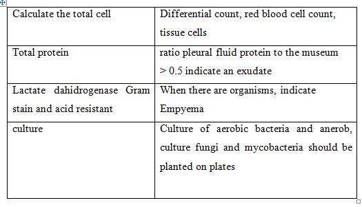
d. bacteriology
Pleural fluid is usually sterile, but can sometimes contain microorganisms, especially when the liquid is purulent, (showing empyema). Purulent effusion which may contain germs that aerobic or anaerobic. Types of bacteria are often found in the pleural fluid is: pneumokok, E. coli, Kleibsiella, Pseudomonas, entero-bacter. On tuberculous pleurisy, the culture fluid of acid-resistant bacteria may indicate a positive hanys to 20%. Laboratory examination of pleural fluid can be seen in the tables below: Laboratory Studies Of Pleural Fluid

7. Bronchoscopy
Bronchoscopy is sometimes done to help find the source of the collected fluid. Bronchoscopy is usually used in cases of neoplasm, corpus alineum in the lung, lung abscess and others
8. Isotope Scanning
Isotope scanning is usually used in cases with pulmonary embolism.
9. Torakoskopi (Fiber-optic pleuroscopy)
Torakoskopi bias is used in cases with pleural neoplasm or tuberculosis. The trick is to do an incision in the chest wall (with a small risk of pneumothorax). Fluid is removed by using suction and air is introduced in order to bias a second look pleura. Using a flexible bronchoscope with a biopsy done.
Medical Management
If the amount of liquid is small, probably only necessary treatment for the cause. If the liquid is much, resulting in suppression or shortness of breath, then periu diiakukan drainage measures (discharge accumulated). Fluid can flow through torakosentesis procedure where a needle inserted into the pleural cavity. Torakosentesis usually performed for diagnosis, but the procedure can also be excluded as much as 1.5 liters of fluid. If the amount of liquid that must be paid more, then put a hose
through the chest wall.
Treatment of acute pleural effusion.
Treatment must consist of the introduction and rapid treatment of primary disease.
A. Efuai require symptomatic pleural fluid drainage pipe with torasentesis or thoracotomy. Not more than 1 to 1.5 L of fluid may be removed at once to prevent pulmonary edema due to reekspansi. When greater numbers to issued, there must be an interval of 1 hour between drainage.
2. Empyema, an exudative effusion is infected, requiring closed tube thoracotomy drainage through okulasi if effusion or pleural caiian if pH less than 7.2.
Treatment of chronic pleural effusion
A. Efusl flowing freely (for example, accompanying the ascitic effusion, parapneumonic effusions with a pH> 7.2) usually resolves after the underlying disease is treated and does not require special treatment.
2. Extensive recurrent effusions accompanying such neoplasms may require repeated drainage with thoracotomy torasentesis or pipe. If the effusion is formed again after 2 or 3 times to do the drainage, should be tried sclerotherapy with chemicals such as tetracycline, 50 mg in 50 mL of salt physiology, or mekloretamin, 10 mg in 50 mL of sterile water.
3. Effusions and empyema requiring drainage berlokulasi. Thoracotomy tube is usually sufficient, but sometimes required surgical drainage.
The handling on:
A. Empyema
In the empyema was given antibiotics and performed spending pus. If nanahnya highly viscous or has accumulated in the fibrous part, the drainage of the pus is more difficult and some of the ribs should be lifted so that it can be installed a larger hose. Surgery is sometimes necessary to cut the layers of pleura terluardari (decortication).
2. Hemotoraks
If blood enters the cavity pleurahempotoraks usually issued through a hose. Through the hose can also be included to help solve the drug blood clots (eg, streptokinase and streptodornase). If bleeding persists or if blood can not be removed through a tube, it is necessary to surgically
3. Kilotoraks
Treatment for kilotoraks performed to repair damage to the lymphatic channels. Can be removed surgically or administration of anticancer drugs to tumors that block the flow of lymph.

Complications Pleural effusion
a. Infections.
Collection of fluid in the pleural space can rrangakibatkan infection (primary empyema), and efus pleura can become infected after the action torasentesis {empyema sekunader). Primary and secondary empyema should be drained and treated with antibiotics to prevent fibrotic reaction. Initial antibiotic selected clinical picture. Choice of antibiotic can be changed after culture results are known.
b. Fibrosis in the lung can be partially reduced by limiting the development of pulmonary ventilation. Fibrotic pleura can also be a source of chronic infection, causing a slight fever.
Pleural decortication, resection by surgery may be required to eradicate infection and restore lung function. Decortication is best done within 6 weeks after the diagnosis of empyema is established, because during this period of the pleural lining is still not well organized (fibrotic) so that the appointment easier.
No comments:
Post a Comment