"A Man can't make a mistake can't make anything"
Showing posts with label symptoms. Show all posts

Monday, 27 August 2012

DEFINITIONS, SYMPTOMS, SIGNS, DIAGNOSIS AND MANAGEMENT ANAL FISTEL/ DEFINISI,GEJALA,TANDA ,DIAGNOSA DAN PENATALAKSANAAN FISTULA ANI


CHAPTER II
Fistula ANI

I. DEFINITION
Fistula ani also called perianal fistula or fistula para-anal1. Anorectal Fistula (Fistula ani) is an abnormal communication between the anus and perianal skin. Glands in the canal lies in linea dentate ani provides a pathway for the infecting organism can achieve intramuscular2 space.

II. Etiology
Fistulas can occur spontaneously or secondary to perianal abscess (or perirektal). In fact, after drainage of the abscess periani, nearly 50% are likely to develop into a chronic fistula. Other fistulas may occur secondary to trauma, Crohn's disease. ani fissures, carcinoma, radiation therapy, aktinomikosis, tuberculosis, and infections klamidia2.
Kriptoglandular hypothesis states that an infection begins in the gland ani and evolve into the muscular wall of the anal sphincter causing anorectal abscess. After surgical or spontaneous drainage periani on the skin, usually of the granulation tissue tract behind, causing symptoms berulang2.
Can be caused by perforation or abscess penyaliran anorektum. Sometimes fistula caused by colitis with proctitis, such as tuberculosis, amubiasis, or morbus Crohn. Infection of the anal glands intersphincter in organisms found in the gastrointestinal tract-either aerobic (Cth: E. coli) and anaerobes (Cth: Bacteroides spp.) - Is a common disorder that causes ini.1

Saturday, 25 August 2012

SYMPTOMS, SIGNS, DIAGNOSIS AND MANAGEMENT oral cancer / GEJALA , TANDA, DIAGNOSA DAN PENATALAKSANAAN KANKER RONGGA MULUT


CHAPTER I
INTRODUCTION

 Diseases of the oral soft tissues has been a serious concern by experts, especially with the increase in cases of deaths caused by cancer in the oral cavity particularly in countries that are developing.
Oral cancer is approximately 5% of all malignancies occurring in men and 2% in women (Lynch, 1994). It has been reported that oral cancer is a major cancer in India, especially in Kerala where the incident was reported at an average height, about 20% of all cancers (Balaram and Meenattoor, 1996).
Although there is progress in the diagnosis and therapy, and death caused by an abnormality of oral cancer is still high and has long been a problem in the world. Some of the reasons put forward for this is mainly due to the lack of early detection and identification of high-risk groups, and the failure to control the primary lesion and cervical lymph node metastases (Lynch, 1994; Balaram and Meenattoor, 1996).
To overcome the problems caused by oral cancer, the WHO has made instructions for oral cancer control, especially for countries that are developing. Control is based on primary prevention where the main principle to reduce and prevent exposure to substances that are carcinogens. The second approach is through the implementation of secondary prevention, in the form of early detection of cancerous lesions and precancerous oral cavity (Subita, 1997). Folson et al, 1972, estimates that 80% of all cases of oral cancer deaths can be prevented with early detection of malignancy in the mouth (Folson et al, 1972).
In general, for the early detection of oral malignancy in the process can be done through anamnesis, clinical examination and confirmed by additional laboratory examinations. In this paper will put forward measures that can be performed by your dentist to detect early malignant processes in the mouth. It is expected to find a dentist lesions suspected of being malignant process early so the prognosis of oral cancer better.


CHAPTER II
Oral cancer

DEFINITION II.1
A. Restriction
Oral cancer is cancer originating from both epithelial mucosa or salivary glands in the walls of the oral cavity and the mouth organ.
 

Wednesday, 22 August 2012

DEFINISI,GEJALA, DIAGNOSA DAN PENATALAKSANAAN HERNIA INGUINAL / DEFINITIONS, SYMPTOMS, DIAGNOSIS AND MANAGEMENT inguinal hernia


BAB I
PENDAHULUAN
Hernia inguinalis merupakan kasus bedah digestif terbanyak setelah appendicitis. Sampai saat ini masih merupakan tantangan dalam peningkatan status kesehatan masyarakat karena besarnya biaya yang diperlukan dalam penanganannya dan hilangnya tenaga kerja akibat lambatnya pemulihan dan angka rekurensi. keseluruhan jumlah operasi di Perancis tindakan bedah hernia sebanyak 17,2 % dan 24,1 % di Amerika Serikat. 1
Hernia inguinalis sudah dicatat sebagai penyakit pada manusia sejak tahun 1500 sebelum Masehi dan mengalami banyak sekali perkembangan seiring bertambahnya pengetahuan struktur anatomi pada regio inguinal.1
Hampir 75 % dari hernia abdomen merupakan hernia ingunalis. Untuk memahami lebih jauh tentang hernia diperlukan pengetahuan tentang kanalis inguinalis. Hernia inguinalis dibagi menjadi hernia ingunalis lateralis dan hernia ingunalis medialis dimana hernia ingunalis lateralis ditemukan lebih banyak dua pertiga dari hernia ingunalis. Sepertiga sisanya adalah hernia inguinalis medialis.Hernia lebih dikarenakan kelemahan dinding belakang kanalis inguinalis. Hernia ingunalis lebih banyak ditemukan pada pria daripada wanita, untuk hernia femoralis sendiri lebih sering ditemukan pada wanita.Sedangkan jika ditemukan hernia ingunalis pada pria kemungkinan adanya hernia ingunalis atau berkembangnya menjadi hernia ingunalis sebanyak 50 % Perbandingan antara pria dan wanita untuk hernia ingunalis 7 : 1. Prevalensi hernia ingunalis pada pria dipengaruhi oleh umur. 1
Hernia merupakan keadaan yang lazim terlihat oleh semua dokter, sehingga pengetahuan umum tentang manifestasi klinis, gambaran fisik dan penatalaksaan hernia penting.

Monday, 23 July 2012

THE ROLE OF SURGERY IN HEMORRHAGIC STROKE


THE ROLE OF SURGERY IN HEMORRHAGIC STROKE

A. DEFINITION

Definition of stroke according to World Health Organization (WHO) is the clinical signs that developed rapidly due to focal brain dysfunction (or global), with symptoms lasting 24 hours or more, and can cause death.
Stroke is a brain attack caused by blockage or sudden onset of rupture of blood vessels of the brain that causes certain brain cells are deprived of blood, oxygen or nutrients and eventually death can occur in these cells in a very short time (Stroke Foundation of Indonesia, , 2006).
Hemorrhagic stroke is rupture of the walls of blood vessels, causing bleeding in the brain. Generally occurs when patients do activities. Bleeding and impairment of consciousness are real (Stroke Foundation of Indonesia, 2006).

B. EPIDEMIOLOGY
Stroke is a major health problem in modern life today. In Indonesia, an estimated 500,000 residents each year occur suffered a stroke, about 2.5% or 125,000 people died, and the remainder mild or severe disability. Number of patients with stroke tend to increase every year, not just attack the elderly, but also experienced by those who are young and productive. Stroke can strike at any age, but which often occurs at the age of 40 years. The incidence of stroke increases with age, the older someone is, the higher the chances of developing a stroke (Stroke Foundation of Indonesia, 2006).
In Indonesia, there are no data to complete epidemiologic stroke, but the proportion of stroke patients from year to year tend to increase. It is seen from the Household Health survey report MOH in various hospitals in 27 provinces in Indonesia. The survey results showed an increase between 1984 and 1986, from 0.72 per 100 patient pada1984 to 0.89 per 100 patients in 1986. In RSU Banyumas, in 1997 stroke patients hospitalized as many as 255 people, 298 people in 1998 sebnyak, in 1999 as many as 393 people, and in 2000 as many as 459 people (Hariyono, 2006).
Stroke or cerebrovascular accident, is the most frequent cause of invalidity in the age group over 45 years in industrialized countries stroke is the third leading cause of death after heart disease and malignancy (Lumbantombing, 1984).

C. Etiology
Hemorrhagic strokes occur because one of the blood vessels in the brain ruptures or tears objec hemorrhagic stroke patients are generally more severe than non-hemorrhagic stroke. Awareness is generally declining. They are in a state of somnolence, osmnolen, spoor, or commas in the acute phase.

D. CLASSIFICATION
According to the cause can be divided into:
1) intracerebral hemorrhage
Intracerebral hemorrhage was found in 10% of all stroke cases, consisting of 80% in the hemispheres of the brain and the rest in the brainstem and cerebellum.
2) Subarachnoid hemorrhage
Subarachnoid hemorrhage is a condition where there is bleeding in the subarachnoid space which arises in the primary.

E. Pathophysiology

Tuesday, 15 May 2012

SURGICAL ASPECT OF DIABETIC FOOT (diagnosis , sign, symptoms, and management ) / komplikasi diabetes melitus pada kaki dan penanganannya.


DIABETIC FOOT SURGERY TREATMENT

A. Definition

Diabetes Mellitus (DM) is a hereditary metabolic disease that mostly, demham signs of hyperglycemia and glucosuria, accompanied by clinical symptoms or absence of acute or chronic, as a result of the lack of effective insulin in the body, lies in the primary disorder of carbohydrate metabolism that is usually accompanied too fat and protein metabolism disorders. (Askandar, 2000).

Gangrene is the process or condition that is characterized by the presence of dead or necrotic tissue, but a microbiological process of necrosis is caused by infection. (Askandar, 2001).

B. Classification

A. Diabetes Mellitus

a. Type I diabetes mellitus (IDDM)

Patients are very dependent on insulin due to an autoimmune process that attacks the insulin. IDDM is the type of DM-derived (Inherited).

b. Type II diabetes mellitus (NIDDM)

Type of DM is influenced by both heredity and environmental factors. A person has a substantial risk of suffering from NIDDM if their parents are people with DM and adopt the wrong lifestyle.

c. Gestational DM

DM of this type tend to occur in pregnant women and the family members who are also suffering from DM. Risk factor is overweight or obese.

d. Secondary DM

DM is associated with other conditions or syndromes (pancreatitis, hormonal abnormalities, and drugs).

Monday, 23 April 2012

BENIGN OR CANCER (MALIGNANT) PAROTID GLAND TUMOR (definition, sign, symptoms, diagnosis, prognosis, and management

Parotid gland


Anatomy of parotid gland
Parotid gland is the salivary glands are paired, numbered 2. Parotid gland is the largest of the salivary glands. Each weighing an average of 25 grams and irregular shape, berlobus, color between green and yellow (Yellowish) located below the external acoustic meatus between the mandible and the muscles sternokleidomastoideus.4

Parotid gland varied shape, when viewed from the lateral triangular 50%, 30% of the top and bottom round. Parotid gland is usually shaped like an inverted pyramid with a surface-surface as follows: superior surface of the small, superficial, anteromedial, and posteromedial. Konkav on the superior surface forms associated with the cartilage of the external acoustic meatus and the posterior part of the temporomandibular joint. Here auriculotemporal nerve supplies the parotid gland. Superficial surface covered by skin and superficial fascia containing a branch of the facial nerve aurikuler, superficial parotid lymph nodes, and the lower limit of platisma.4
The anterior part of the gland adjacent to the posterior edge of mandibular ramus and slightly coat the posterior edge of masseter muscular. Posterior part of the gland is surrounded by the ear, mastoid processus, and the anterior edge of the muscular stemokleidomastoideus. The inside of the medial lobe extends into the cavity parafaring, limited by the processus stilomandibular stiloideus and ligament, muscular digastrikus, and carotid sheath. In the anterior lobe is located adjacent to the medial ptetygoideus. Lateral parts covered only by skin and subcutaneous fat tissue. Connective tissue and fat tissue from the fascia of the neck in this gland wraps. Parotid gland is closely linked to important structures around the internal jugular vein and its branches, the external carotid artery and its branches, lymph glands, auriculotemporalis of nerve branches and nerve trigerninus fasialis.4

Tuesday, 17 April 2012

ORAL-ONCOLOGY SURGERY SERIES : ORAL CAVITY CANCER / kanker rongga mulut ( definition, sign, symptoms, etiology, diagnosis and management)


CHAPTER I
INTRODUCTION

Diseases of the oral soft tissues has been a serious concern by experts, especially with the rising cases of deaths caused by cancer in the oral cavity particularly in countries that are developing.
Oral cavity cancer is approximately 5% of all malignancies occurring in men and 2% in women (Lynch, 1994). It has been reported that oral cancer is the leading cancers in India especially in Kerala where the incident was reported at an average height, about 20% of all cancers (Balaram and Meenattoor, 1996).
Although there are developments in the diagnosis and therapy, abnormalities and mortality resulting from oral cancer is still high and has long been a problem in the world. Several reasons are put forward for this is mainly due to the lack of early detection and identification of high risk groups, as well as failure to control the primary lesion and cervical lymph node metastasis (Lynch, 1994; and Meenattoor Balaram, 1996).
To overcome the problems caused by oral cancer, WHO has made instructions to control oral cancer, especially for countries that are developing. Control is based on primary prevention measures where the main principle to reduce and prevent exposure to substances that are carcinogens. The second approach is through the implementation of secondary prevention, ie early detection of cancerous lesions and precancerous oral cavity (Subita, 1997). Folson et al, 1972, estimates that 80% of all cases of oral cancer deaths can be prevented with early detection of malignancies in the mouth (Folson et al, 1972).
In general, for the early detection of malignancies in the mouth can be done through anamnese, clinical examination and confirmed by additional tests in the laboratory. In this paper will put forward measures that can be done by the dentist to detect early malignant processes in the mouth. It is expected to find a dentist suspected lesions as malignant process early so that the prognosis of oral cancer is better.

CHAPTER II
Oral cavity cancer

II.1 DEFINITIONS
A. Restriction
Oral cavity cancer is cancer that originates in either coming from the mucosal epithelium or the salivary glands in the oral cavity wall and organs in the mouth.
   

Friday, 13 April 2012

PAEDIATRIC SURGERY SERIES : INVAGINASI/ INTUSSUCEPTION (sign, symptoms, diagnosis and management)


CHAPTER I
INTRODUCTION

I.1 Background

Invagination events often occur in the rainy season in obese children who have flu and diarrhea. invagination is an acute intestinal peristaltic disorders, in which a segment of intestine protrudes into the next segment. Generally the proximal segment into the distal segment. (1) invagination is often found in children and rare in young people and adults.
Invagination in children and infants is still difficult to find compared with invagination in adults. Invagination in children and infants is often found in under 2 years of age and most found at the age of 5-9 months. The cause of invagination in children and infants 70% -90% is unknown; some literature linking the hypertrophied Peyer's patches caused by a virus infection, weather changes or changes in eating patterns. While a large invagination in children and adults the cause is a pathological disorder (Meckel diverticular, polyps, tumors). On the other references say 70% of patients under 1 year (often at the age of 6-7 months). The incidence varies from 1-4 per 1000 live births, and four times more men than women. As well as more common in babies with good nutrition. (1.2)
Invagination cases admitted to hospital as emergency cases. The first action is to overcome the lack of fluid, electrolyte and acid-base balance.

Thursday, 12 April 2012

UROLOGY / PAEDIATRIC SURGERY SERIES : WILM'S TUMOR/ NEPHROBLASTOMA. ( Definition, etiology, stadium, sign, symptoms, diagnosis, and management)

Wilm Tumor (Nephroblastoma)

A. INTRODUCTION

Definition of Wilm tumor is malignant and the most to the five most common renal tumor at the age of the children. These tumors appear most at the age of three years and is rarely found after 8 years of age. There are 250 cases of Wilm tumor ever reported. However, preoperative diagnosis of Wilm tumor in adults is quite difficult. These tumors usually appear as a solid renal mass with or without accompanied by hematuria. This solid mass may be accompanied by the process of cystic degeneration and focal necrosis. Sometimes the radiological picture may resemble renal cysts hemorhagik. These tumors appear in one or two kidneys. (A) (7)

The existence of large masses diabdomen, especially in children aged 1-5 years should arouse suspicion Tumor Wilm. This is an aggressive neoplasm and metastasis to different organs, but to respond to combination therapy. Wilm tumor when diagnosed and get appropriate therapy has a high cure rate is as high as 90%. (A) (7)

Development of diagnostic imaging in recent years also plays an important role in enhancing the efficacy and accuracy of diagnosis of Wilm tumor and it is extremely important in designing the best combination therapy in children. (A) (7)

BASIC KNOWLEDGE ABOUT THYROID GLAND AND aberrant thyroid / THYROID ACCESSORIES / ECTOPIC. (SIGN, Symptoms, Etiology, DEFINITION, DIAGNOSIS AND MANAGEMENT). PENGETAHUAN DASAR TENTANG KELENJAR THYROID DAN THYROID ABERRANT / AKSESORIS / EKTOPIK


The thyroid gland
The thyroid gland is one of the largest gland, which normally weighs 15 to 20 grams. Thyroid gland excrete  three kinds of thyroid hormones, thyroxine (T4), triiodothyronine (T3), and calcitonin.


Figure 1. Thyroid anatomy
In anatomy, the thyroid is an endocrine gland (ductus have not) and bilobular (right and left), connected by the isthmus (bridge) located in front of the trachea just below the cartilago cricoidea. Sometimes there are additional lobes that extends into the upper (ventral body), the lobes of the pyramid.
In embryology, the stage is the formation of the thyroid gland:
• The thyroid gland was originally a two protrusions of the front wall of the middle farings, which is formed at the age of 4 weeks of birth. The first protrusion called the pharyngeal pouch, which is between 1 and 2 brachial arch. The second protrusion on ceacum foramen, which is under the ventral branch farings I.
• At week 7, the protrusion of the foramen caecum pharyngeal pouch will go through a channel called the ductus thyroglossus.

Wednesday, 11 April 2012

TERATOMA AND DERMOID CYST, DEFINITION, SIGN, SYMPTOMS, ETIOLOGY, DIAGNOSIS AND MANAGEMENT


CHAPTER I
INTRODUCTION

I. 1 OVERVIEW
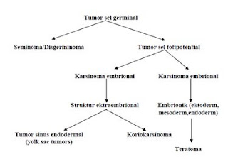
Teratomas are germ cell tumors are generally composed of multiple cell types derived from one or more of the three germ layers of endoderm, mesoderm and ectoderm. Inconsistent nomenclature is often confused about the various subtypes of teratoma. Teratomas derived from the Greek meaning of the terrace monster, which was first described by Virchow in the first edition of his book published in 1863. Teratoma is divided into three categories: mature teratoma (benign), immature teratoma and teratoma with differentiation monodermal particular degrees depending on the quantity of immature tissue showed the potential for malignancy. Cystic teratomas are generally benign and malignant solid is. Teratomas vary from a benign form of the cystic lesions of well differentiated (mature) to form a solid and malignant (immature). Immature teratomas are malignant germ cell tumor to the three most common after disgerminoma and endodermal sinus tumor. In addition, there also have a specific component (usually squamous) who experienced malignant transformation, but are rarely found.

In 1831, Leblanc created the term dermoid cyst in the literature of veterinary medicine when he succeeded in removing skin lesions at the base of the skull resembles a horse. Both teratomas and dermoid, a term still used and are often used interchangeably. Implications of these elements behawa beginning to resemble the skin with a complementary structure composed of dermoid, whereas teratoma has no such limit. Dermoid now known as the trigeminal and contain various types of tissue.

Monday, 9 April 2012

ONCOLOGY / ORTHOPAEDI SURGERY SERIE : SOFT TISSUE CANCER / MALIGNANT FIBROUS HISTIOCYTOMA ( definition, signs, symptoms, diagnosis and management)

INTRODUCTION

In America, more than 2000 people are diagnosed with this tumor each year, mostly one type of cancer suffered by children and adolescents, but few cases are found in adults.
      Cancer around the bone itself consists of several types, namely osteosarcoma, Ewing's sarcoma, chondrosarcoma, malignant fibrous histiocytoma, fibrosarcoma, and Chordoma. Malignant fibrous histiocytoma (MFH) is a soft tissue sarcomas of the most frequently in older adulthood. There are several variants of the MFH, storiform-pleomorphic subtype, myxoid, inflammatory and giant cell. Infammatory and giant cell is a rare subtype. Most MFH tumors found in the soft tissue in the extremities and body. Giant cell MFH found in older patients.

CHAPTER I
DISCUSSION

I.1 Definition
Malignant fibrous Histiositoma (malignant fibrous histiocytoma) is a sarcoma that usually begins in the soft tissues. This condition usually appears as a large mass that is painful and can lead to fractures due to bone destruction by tumor spread. 
Malignant fibrous histiocytoma was first introduced in 1961 by Kauffman and Stout. They describe as a tumor rich in histiocytes MFH with storiform growth pattern. In 1977, MFH is considered as soft tissue sarcomas of the most common of adult life.

I.2 Epidemiology
Malignant fibrous histiocytoma of soft tissue usually appears in patients is approximately 50 to 70 years although it can occur at any age. Malignant fibrous histiocytoma is very rare in people less than 20 years. More men than in women. 
Malignant fibrous histiocytoma has a 44% recurrence rate and metastasis rate by 42%. Incidence of metastases to regional lymph nodes are found as much as 12%. 

I.3 Histiologi
      Histologist is divided into four subtypes:
A. Storiform-pleomorphic
  Of these, storiform-pleomorphic is the most common type, accounting for up to 70% of cases, see Figure 1.

Figure 1: A histologic specimen shows fibrous histiocytoma classic storiform-pleomorphic malignant. Microscopically, the pattern shows fasikula short storiform spindle cells radiating from a central point which is mixed with giant neoplastic cells in the pleomorphic

2. Myxoid
Figure 2: Example of histological type myxoid MFH. So that the tumor will be marked as a variant of myxoid, myxoid tissue must explain at least half of the tumor

Friday, 6 April 2012

PLASTIC / Oncologic Surgery SERIES: THE DISTINGUISHING Bell's palsy with Iatrogenic paralise of the facial nerve (etiology, SIGN-symptoms, DIAGNOSIS AND MANAGEMENT)


PLASTIC / Oncologic Surgery SERIES: THE DISTINGUISHING Bell's palsy PARALISE iatrogenic facial nerve (etiology, SIGN-symptoms, DIAGNOSIS AND MANAGEMENT)


The term Bell's palsy is a peripheral facial nerve paresis of unknown cause (idiopathic) and acute. Many are mixed up between Bell's palsy with peripheral facial nerve paresis other unknown causes.
Bell's palsy is found, usually people with facial paralysis learn from friends or family or at the mirror or brush teeth / rinsing. When the patient realizes that he is paralyzed on her face, then he began to feel fear, shame, low self-esteem, cosmetic and sometimes disturbing soul depressed, especially in women and in patients who have a profession that requires him to appear in public. Often the question arises in his heart, if his face could go back to normal or not.

from wikipedia picture
 from freedictionary.com

Tuesday, 27 March 2012

MALIGNANT SKIN TUMOR / SKIN CANCER AT AGLANCE ( etiology,sign, symptoms,diagnosis and management of Basal Cell Carcinoma, Squamous Cell Carcinoma,and Malignant Melanoma )

 Malignant SKIN TUMOR AT AGLANCE
  ( Basal Cell Carcinoma, Squamous Cell Carcinoma, Malignant Melanoma )

Definition
Malignant skin tumor is a disease characterized by the growth of skin cells that are not controlled, can damage surrounding tissue and can spread to other parts of the body. Because the skin consists of several types of cells, the skin cancer also vary according to cell type affected.

Epidemiology
Adult skin cancer tend to have increased in number especially in the Americas, Australia and Britain. Based on several studies, those white people are more likely to suffer this type of skin cancer. It is predicted as a result of their frequent exposure (lots of exposure) sunlight. In Indonesia people with skin cancer is fairly small compared to the third-countries, however, skin cancer needs to be understood because in addition to causing defects (damage the appearance) is also at an advanced stage can be fatal to the patient (1).
Malignant skin tumor types that are found throughout the world are basal cell carcinoma (basalioma), squamous cell carcinoma, a relatively non melanoma and malignant melanoma. Basal cell carcinoma is the most common. In America, about 800,000 people suffering this cancer every year. 75% of skin cancer are basal cell kanser. Squamous cell carcinomas are found to be what the 200,000 Americans each year. Melanoma is the most rare but cause the most deaths. According to WHO, as many as 160,000 people  suffering melanoma each year and as many as 48 000 deaths are reported each year (3).

Skin Embryology (4)
The skin has a double origin:
(A) the superficial layer (epidermis), formed from the surface ectoderm.
(B) inner layer (dermis), derived from the underlying mesenchyme.

A. Epidermis
At first, the embryo diilapisi by a single layer of ectoderm cells (Fig. 20.1A). At the beginning of the second month, the epithelium is split and formed a flat layer of cells, or epitrikium periderm, on its surface (Figure 20.1B). In the subsequent cell proliferation in the basal layer, forming a third zone (intermediate zone) (Figure 20.1C). Finally, at the end of the fourth month, the epidermis obtain definitive arrangement, and can be recognized four layers (Fig. 20.1D)


Thursday, 8 March 2012

Etiology,sign, symptoms,Diagnosis and management Benign Prostat Hyperplastic (hyperplasia / hypertrophy)

Introduction
Enlargement of the prostate gland has a significant morbidity in a population of elderly men. The symptoms are a common complaint in the field of urologic surgery.
Prostate hyperplasia is one of the major health problems for men over the age of 50 years and was instrumental in the decline in quality of life. A study says that one third of men aged between 50 and 79 years of experience prostatic hyperplasia.
Exact prevalence in Indonesia is not yet known but is estimated based on foreign literature since age 50 years 20% -30% of patients will require treatment for prostatic hyperplasia. Which obviously depends on the prevalence of child bearing age. Actually, the changes towards the enlargement of the prostate is started early, beginning in mikroskopoik changes which then manifests into macroscopic abnormalities (enlarged gland) and then a new clinical symptoms.
Based on the autopsy rate is microscopic changes in the prostate can be found at the age of 30-40 years. When these microscopic changes constantly evolving there will be changes anatomic pathology. In men aged 50 years and the number of events around 50%, and at the age of 80 years about 80%. Approximately 50% of the numbers mentioned above will cause the symptoms and clinical signs.
The existence of this hyperplasia would lead to urinary tract obstruction and to overcome this obstruction can be done in various ways ranging from the most minor of action that is a conservative (non operative) to the most severe action that is operating.
I. ANATOMY AND PHYSIOLOGICAL
Under normal circumstances the prostate is approximately the size of a walnut. Normal prostate weight in adults is ± 20 grams. Located around the prostatic urethra and bladder neck as well as between the urogenital diaphragm. The apex of the prostate is located on the external urethral sphincter of the bladder. In the anterior symphysis pubis adjacent to but separated by a cavity in the extraperitoneal fat retopubis (Retzius cavity). In the posterior prostatic fascia is separated from the rectum by denonvilliers.


Thursday, 23 February 2012

Hydrocele and hernia at a Glance (etiology,sign, symptoms,diagnosis and management)


A.      ANATOMY
Generally hernia is a protussion the content of the cavity through  defect or the weakness of the cavity wall in question. On abdominal hernia, abdomenal content protunding  through a defect or weakness of the musculo-aponeurotik layer of the abdominal wall. Based on the occurance, congenital hernia or hernia are divided into congenital and acquired hernia. Hernia are named according to its location, eg diaphragm, inguinal, umbilical and femoral


By their very nature, can be called a hernia hernia when the hernia contents can reponibel jeluar entry. Out when standing or straining, and came back when sleeping or pushed into the stomach. When the contents of the bag can not be repositioned back into the abdominal cavity, called a hernia hernia ireponibel. This is usually caused by adhesions in the peritoneal pouch bag hernia. This is called a hernia hernia accreta. There is no pain or signs of intestinal obstruction.

Inkarserata called hernia or hernia strangulate when it squashed by the hernia ring so that the bag can not be caught and returned to the abdominal cavity. As a result, frequent passage or vascular disorders. Inkarserata hernia clinically more intended for hernia ireponibel with passage disturbance, whereas vascular disorder known as Strangulated hernia.
External hernia is an abnormal protrusion of intra-abdominal organs through the abdominal wall defect in the fascia. Hernia which often happens is inguinal, femoral, umbilical, and paraumbilikal.

Inguinal hernia is the protrusion viscus (organ) from the peritoneal cavity into the inguinal canal.

All hernia occurs through slit weak or potential weakness in the abdominal wall that is triggered by an increase in intra-abdominal pressure that repeated or continuous.


Seventy-five percent of all abdominal hernias occur in the inguinal (groin). Others may occur in the umbilicus (navel) or other abdominal area. Inguinal hernias are divided into two, namely the medial inguinal hernia and inguinal hernia lateralis. If the bag reaches the lateral inguinal hernia scrotum (testicles), called a hernia hernia skrotalis. Lateral inguinal hernia occurs more frequently than the medial inguinal hernia with a ratio of 2:1, and among men it was 7-fold more frequently affected than women. The more we age, the greater the possibility of a hernia. This is influenced by the strength of abdominal muscles that have been weakening.

Hernias that arise in the crease and the inguinal hernia abdominokrural is arising below the fold is a femoral hernia. Inguinal canal is an oblique line passing through the lower anterior abdominal wall. This channel allows the structures to pass to and from the testis to the abdomen in the male. In women, the channel is crossed by ligaments rotundum uteri, magi from the uterus to the labium. In addition, the channel is bypassed Ilioinguinalis nerve in both sexes.
            The length of the inguinal canal in adults is approximately 4 cm, formed from the profundus inguinal annulus / annulus inguinali internal to the superficial / external. Inguinal canal is located parallel to and just above the inguinal ligament. In neonates, the internal inguinal annulus is located almost directly posterior to the external inguinal annulus so that the inguinal canal is very short at this age. Then, the internal annulus moves toward the lateral growth effect.
            Internal inguinal annulus is an oval hole in the fascia transversalis, located about 3 cm above the inguinal ligament, midway between the Messiah and symphisis pubis.Medial to the internal annulus there is av. inferior epigastric. Edge of the annulus is the origin of the internal spermatica fascia in the male or the inner wrapper rotundum uteri rotundum ligaments in women.
            Externa is an annular shaped defect in the inguinal triangle (Hesselbach's triangle) on the aponeurosis. Obliquus externus abdominis and essentially formed by the crista pubica. Edge of the annulus is the origin of the fascia spermatica externa. Lateral boundary is the inferior epigastric artery, the medial border of the m. rectus abdominis lateral part, and the inferior border of the inguinal ligament.
            Inguinal canal is formed on the wall of the anterior, posterior, superior, and inferior.Anterior wall formed by the aponeurosis m. Obliquus externus abdominis is amplified at 1/3 by the lateral fibers of m. Obliquus internus abdominis. The entire length of the posterior wall of the inguinal canal is formed by the fascia transversalis tendon reinforced cojoint in 1/3 medial. Cojoint tendon is the tendon insertion combined m. Obliquus internus abdominis and m. transversus abdominis is attached to the crista pubica and linea pectinea. Basic or inferior wall of the inguinal canal is formed by the inguinal ligament, while the roof is formed by m. Obliquus abdominis internus abdominis and m.transversus.

Figure 1. Hesselbach's triangle
Inguinal hernias can be directly (direct) and can also indirectly (indirect). Indirect inguinal hernia bag of walking through the deep inguinal ring, lateral to the inferior epigastric vessels, and finally towards the scrotum. Pockets of directors inguinal hernia protruding directly through the base of the inguinal canal, medial to the inferior epigastric vessels, and rarely go down towards the scrotum. Femoral hernia is almost always seen as a mass irredusibel, although pockets more kososng, because fat dam of femoral canal lymph nodes around the bag. Single, enlarged lymph nodes can mimic femoral hernia very quickly.
Indirect hernia bag is actually a process vaginalis persistently dilated. Hernia was walking through the deep inguinal ring and follow the sheath into the scrotum. At the deep ring, bag filling anterolateral side of the cord. Properitoneal fat pockets often associated with indirect and is known as a lipoma of the cord, although the fat is not a tumor.
Retroperitoneal organs such as the sigmoid colon, cecum and ureter can be slipped into a pocket indirect. In the bag, these organs become part of the pouch wall and are susceptible to injury during repair.
Inguinal hernia pouch directors come from the bottom of the inguinal canal, the Hesselbach triangle; protruding hernia bag directly and does not contain obliqus externus muscle aponeurosis. Only in rare circumstances, the hernia is so intense that it pushes out through the annulus superficial and descend into the scrotum. The bladder is often a component of the bag is empty hernia director.
Bags from the femoral hernia femoral canal through a defect on the medial side of the femoral sheath (femoral sheath). Femoral canal contains one or two lymph nodes, which are scattered called Cloquet. Lymph-nodes are pushed out of the femoral canal by a protrusion of peritoneal and often form a palpable mass.


Phylloid Tumor (Mamae) / Cystosarcoma phylloides Mamae ( etiology,sign, symptoms,diagnosis and management)


                                                                     CHAPTER I
                                                                INTRODUCTION

It is a benign neoplasm that is locally infiltrate and possibly malignant (10-15%). Used to be called Cystosarcoma phyllodes are benign tumors that occur almost exclusively on women's breasts. Its name comes from the Greek word sarcoma, which means the fleshy tumor, and phyllo, meaning leaf. So called because the tumors display characteristics of a large, malignant sarcomas, such as display-leaves when cut, and the epithelium, cyst-like space when viewed histologically. Because most of the tumor was benign tumor is also called filoides.
Although metastatic tumors are not benign, but they have a tendency to grow locally aggressive and recurrent. Similar to sarcoma, malignant tumor hematogenous metastasis. Filoides tumor pathological picture is not always predict the clinical behavior of neoplasms, hence in some cases there is uncertainty about the classification level of the lesion.
The characteristics of malignant tumor filoides are as follows:
· Recurrent malignant tumors are more aggressive look than the original tumor.
· Pulmonary metastasis is the place most frequently, followed by bone, heart, and liver.
· The symptoms for metastatic involvement can occur immediately, until a few months and no later than 12 years after initial therapy.
· Most patients with metastases died within 3 years of initial therapy.
· There is no treatment for systemic metastases occurred.
· Roughly 30% of patients with malignant tumors filoides died of the disease.

            Filoides tumor etiology is not known, it is estimated associated with fibroadenoma in some cases, because patients may possibly have a second histological lesions in the same tumor. However, if the tumor develops filoides of developing fibroadenoma or both together, or whether the tumor can arise de novo filoides, is still unclear
This tumor has a smooth texture, sharply bounded and usually move freely. This tumor is relatively large tumor, with an average size of 5 cm. However, lesions> 30 cm have been reported.
Haagensen reported roughly one for every 40 filodes tumor fibroadenoma. Distribution of age according to Haagensen majority occur between the ages of 35 and 55 years.Bilateral tumors are very rare and rarely also occur in patients under age 20, first appeared benign react.
Mammography and ultrasound examination (Figure II.03) are generally important in the diagnosis of breast lesions, but they are notoriously unreliable in differentiating benign tumor from filoides malignant form of the condition or of fibroadenoma. Thus, the findings on imaging studies is not a definite diagnosis. Open excision biopsy of breast lesions smaller or incisional biopsy for larger lesions is a surefire method for diagnosing tumor filoides.

 image 03. Mammography image of the tumor filoides