"A Man can't make a mistake can't make anything"
Showing posts with label sign. Show all posts

Monday, 23 July 2012

diagnosis, sign, symptom, and management brain gliobastoma


GLIOBASTOMA
                            
INTRODUCTION
Number of primary CNS neoplasms estimated 9% of all primary tumor types. Intracranial neoplasms arising in an estimated 85% of cases. In general, primary brain tumor is a type of neuroglial., These tumors arise from the parenchyma and are generally derived from glioma. Approximately 6600 new cases of malignant glioma are reported each year. Meningioma is the second largest group of intracranial happens, roughly around 15%. The pituitary gland is the most frequently as the tumor growth, usually the type of adenoma. Roughly about 10% of all intracranial tumors. Neoplasm metastases are common. The incidence depends on age, and usually occurs after the fourth decade. An estimated 1/6 of brain tumor metastases will occur no symptoms. Treatment of tumors depends on tumor type, clinical condition, the need for therapy before surgery, and the handling time of surgery and post surgery. The most common glioma is a tumor.

DEFINITION
Glioma is a tumor originating from the brain tissue. Glioma is a tumor that can invade penginflitrasi some parts of the brain. Malignant gliomas usually occur in many brain neoplasm whose number is about 45% of all brain tumors. Usually these tumors can not be removed completely, because the tumor spreads by infiltration into the surrounding nervous tissue and it is not considered to resect completely without causing damage to vital structures.

Sunday, 15 July 2012

DIAGNOSIS,SIGN ,SYMPTONS AND MANAGEMENT BREAST CANCER

CHAPTER I
INTRODUCTION

 Breast tumors often give the impression of scary, especially when found in women aged over 40 years. Even many of the oncologists argue that any tumor in the breast carcinoma, especially in women considered at high risk groups such as fibroadenoma, although benign tumors, dysplasia (fibrocystic, adenosis), mastitis and fat necrosis can not be ignored. This opinion is understandable, given the frequency of breast carcinomas are relatively high, giving rise to the problem health dala especially for women, not only in developed countries, but also in developing countries including Indonesia. The frequency of breast carcinoma in developed countries is the most that is the ratio of 5:1, compared with carcinoma of the cervix uteri, while in Indonesia ururtan neoplasms are located in the second after the cervix uteri karsinomaa.


CHAPTER II
Anatomy and Physiology

I. ANATOMY OF BREAST
   The stroma of breast masses and breast parenchyma located in the anterior wall of the piston between the ICS II and VI and parasternal line to axilaris medius. Breasts consist of alveoli, lactiferous duct, lactiferous sinus, ampulla, pore pailla, and alveola edge. The main vascularization of the breast have a branch. the internal mammary, a. torakoakromialis and a branch. Intercostal.
Breast lies on either side hemitoraks with limits as follows:
A. Boundaries of the breast that looks from the outside:
a. Superior: ribs II or III
b. Inferior: VI or VII ribs
c. Medial: the edge of the sternum
d. Lateral: anterior axillary line
2. The boundaries of real breasts:
a. Superior: almost to the clavicle
b. Medial: midline
c. Lateral: m. latissimus dorsi

Tuesday, 15 May 2012

SURGICAL ASPECT OF DIABETIC FOOT (diagnosis , sign, symptoms, and management ) / komplikasi diabetes melitus pada kaki dan penanganannya.


DIABETIC FOOT SURGERY TREATMENT

A. Definition

Diabetes Mellitus (DM) is a hereditary metabolic disease that mostly, demham signs of hyperglycemia and glucosuria, accompanied by clinical symptoms or absence of acute or chronic, as a result of the lack of effective insulin in the body, lies in the primary disorder of carbohydrate metabolism that is usually accompanied too fat and protein metabolism disorders. (Askandar, 2000).

Gangrene is the process or condition that is characterized by the presence of dead or necrotic tissue, but a microbiological process of necrosis is caused by infection. (Askandar, 2001).

B. Classification

A. Diabetes Mellitus

a. Type I diabetes mellitus (IDDM)

Patients are very dependent on insulin due to an autoimmune process that attacks the insulin. IDDM is the type of DM-derived (Inherited).

b. Type II diabetes mellitus (NIDDM)

Type of DM is influenced by both heredity and environmental factors. A person has a substantial risk of suffering from NIDDM if their parents are people with DM and adopt the wrong lifestyle.

c. Gestational DM

DM of this type tend to occur in pregnant women and the family members who are also suffering from DM. Risk factor is overweight or obese.

d. Secondary DM

DM is associated with other conditions or syndromes (pancreatitis, hormonal abnormalities, and drugs).

Wednesday, 2 May 2012

TRAUMATOLOGY SERIES : UPDATED ABDOMINAL TRAUMA AT AGLANCE. (definition, diagnosis, clinical overview, sign, symptom, and management )

ABDOMINAL TRAUMA

CHAPTER I
INTRODUCTION


I. Anatomy of the abdomen

I. 1 Anatomy of the external abdominal

a. Front abdominal
Front abdominal definition is a restricted area in the superior by intermamaria line, in inferior limited by the inguinal ligament and lateral to the pubic symphysis and both anterior axillary line.

b. Waist
Is an area that lies between the anterior axillary line and posterior axillary line, from between the ribs to 6 above, down sampaicrista iliaca. At this location there is a thick wall of the abdominal muscles, as opposed to the thinner wall of muscle on the front, especially tterhadap menjadipelindung stab wounds.

c. Back
This area is located in the back of the posterior axillary line, from the lower end of the scapula to the crista iliaca. As with daerak flank, here back muscles and paraspinal muscles to protect against sharp trauma. (1)


I. 2 Anatomy of the abdomen in


There are three rooms, the peritoneal cavity, retroperitoneal cavity and pelvic cavity. The pelvic cavity contains the parts of the peritoneal cavity or retroperitoneal cavity.

a. Peritoneal cavity
Simply the peritoneal cavity is divided into two parts, the top and bottom. The peritoneal cavity is protected by the bottom of the thoracic wall which includes the diaphragm, liver, spleen, gastric and transverse colon. This section is also called a thoracoabdominal components of the abdomen. At the time of the diaphragm rises up between the ribs IV at full expiration, each rib fracture or penetrating stab wound below the intermamaria can injure organs in the abdomen. The peritoneal cavity contains the small intestine, the colon ascendens and colon descendens, sigmoid colon, and in women, internal reproductive organs.


b. Retroperitoneal cavity

Potential cavity is a cavity behind the peritoneum lining the abdominal wall, and includes the abdominal aorta, inferior vena cava, a large part of duodenoum, pancreas, kidney and ureter and the posterior part of the colon ascendens and colon descendens, and also the retroperitoneal pelvic cavity. Injury to the retroperitoneal organs are difficult to identify because this area is far from the reach of regular physical examinations, and also here in the first injury will not show any signs or symptoms of peritonitis. In addition, the cavity is not included in the sample examined in diagnostic peritoneal lavage (DPL).

c. The pelvic cavity

Pelvic cavity, which is protected by the bones of the pelvis, is actually the bottom of the intraperitoneal cavity, while the bottom of the retroperitoneal cavity. Contained therein rectum, vesica urinary, iliaca vessels, and in women, internal reproductive organs. As with the thoracoabdominal, examination of pelvic organs obstructed by parts of the bones on it. (1)

I. Regio-3 region of the abdomen

The abdomen is divided into nine regions, namely:

1. Epigastrica Regio (right and left)
2. Hipocondria Regio (right and left)
3. Umbilical region (right and left)
4. Region of the lateral (right and left)
5. Pubica Regio (right and left)
6. Inguinal region (right and left)

Monday, 30 April 2012

STANDARD TREATMENT OF PATIENTS HIV / AIDS (acquired immune deficiency syndrome), WHEN HAVE SURGERY. STANDAR PENANGANAN TERHADAP PENDERITA HIV / AIDS, KETIKA MENGALAMI PEMBEDAHAN (DEFINITION, SIGN, SYMPTOMS, DIAGNOSIS, PROGNOSIS AND MANAGEMENT)


STANDARD TREATMENT OF PATIENTS HIV / AIDS, WHEN HAVE SURGERY

CHAPTER I
INTRODUCTION

HIV infection is a viral infection that progressively destroys white blood cells and causes AIDS (Acquired Immunodeficiency Syndrome). (10) AIDS was first discovered in 1981. (1)
Transmission of HIV infection from mother to child is the main cause of HIV infection in children under 15 years of age. Since HIV became pandemic in the world, an estimated 5.1 million children globally are infected with HIV. Most of these patients infected through transmission from mother to child. Each year an estimated more than 800,000 infants become infected with HIV through transmission from mother to child. And followed by the approximately 610 000 child deaths from the virus. (10)
            In Indonesia, according to the Directorate General of Health Department of PPM and PL recorded 3568 cases of HIV / AIDS at the end of December 2002. There are 20 children with HIV infection who are infected mother. Research conducted Pelita Science Foundation and The Faculty of medicine obstetrics / RSCM during the years 1999-2001 do a check on 558 pregnant women in poor areas in Jakarta, showed as many as 16 people (2.86%) suffering from HIV infection. (10)
Women are contracting HIV infection through heterosexual relations with an infected partner or through the use of drugs, increasing HIV infection in children is due to the result of transmission during the perinatal period (pregnancy, during and after childbirth). Perinatal HIV transmission is a major cause of pediatric AIDS mother - mothers that transmit HIV are usually good and has a number of CD 4 T lymphocytes are normal and do not know if they are infected with HIV. More than 90% of AIDS in children were reported in 1994 occurred because of transmission from mother to child. (10)
 Transmission to the infant can occur during pregnancy, delivery or postnatal through breast milk. The incidence of mother to child transmission is estimated at 20% - 30%. HIV transmission to the fetus if no intervention reported to range between 155-45%. (10)
The risk of transmission in developing countries around 21% - 43%, higher than the risk of transmission in developed countries about 14% -26%. Transmission can occurs during pregnancy, intrapartum, and postpartum. The risk of transmission of most infections occur during labor by 18%, in the content of 6% and 4% after delivery.


Thursday, 26 April 2012

MY HEAD ENLARGED AND MY SUN SET EYES : HYDROCEPHALUS (definition, anatomy, diagnosis, sign, symptom, and management)


CHAPTER I
INTRODUCTION

Hydrocephalus comes from hydro meaning water and chepalon which means head. Hydrocephalus is a buildup of cerebrospinal fluid (CSS) is actively causing dilatation of the ventricular system of the brain where there is excessive accumulation of CSF in one or more of the ventricles or subarachnoid space. This situation is caused because there is imbalance between production and absorption of the CSS. If the excessive accumulation of CSF occurs on the cerebral hemispheres, a condition called subdural higroma or subdural fluid collection. In the case of excessive fluid accumulation occurs in the ventricular system, a situation known as hydrocephalus internal.Selain that some intracranial lesions causing elevation of ICT, but not until the cause of hydrocephalus. CSS is not equivalent to raising the volume with hydrocephalus; it also occurs in cerebral atrophy.


Hydrocephalus as a clinical entity distinguished by three factors: a). Intraventrikuler pressure elevation, b). Addition of CSS volume, c). Dilatation of the cavity CSS. Overall, the incidence of hydrocephalus is estimated close to 1: 1000. while the incidence of congenital hydrocephalus varies for each of the different populations. BL Hershey said most of hydrocephalus in children is congenital is usually seen in infancy. If hydrocephalus appears after age 6 months is usually not because of congenital. Mujahid Anwar et al get 40-50% of infants with intraventricular hemorrhage grade 3 and 4 had hydrocephalus. Pongsakdi Visudiphan et al in a study 36 of 49 children with TB meningitis had hydrocephalus, with a second note 8 children with obstructive hydrocephalus and 26 children with communicating hydrocephalus. Hydrocephalus that occurs as a complication of bacterial meningitis can be found at any age, but more often in infants than children.

Monday, 23 April 2012

BENIGN OR CANCER (MALIGNANT) PAROTID GLAND TUMOR (definition, sign, symptoms, diagnosis, prognosis, and management

Parotid gland


Anatomy of parotid gland
Parotid gland is the salivary glands are paired, numbered 2. Parotid gland is the largest of the salivary glands. Each weighing an average of 25 grams and irregular shape, berlobus, color between green and yellow (Yellowish) located below the external acoustic meatus between the mandible and the muscles sternokleidomastoideus.4

Parotid gland varied shape, when viewed from the lateral triangular 50%, 30% of the top and bottom round. Parotid gland is usually shaped like an inverted pyramid with a surface-surface as follows: superior surface of the small, superficial, anteromedial, and posteromedial. Konkav on the superior surface forms associated with the cartilage of the external acoustic meatus and the posterior part of the temporomandibular joint. Here auriculotemporal nerve supplies the parotid gland. Superficial surface covered by skin and superficial fascia containing a branch of the facial nerve aurikuler, superficial parotid lymph nodes, and the lower limit of platisma.4
The anterior part of the gland adjacent to the posterior edge of mandibular ramus and slightly coat the posterior edge of masseter muscular. Posterior part of the gland is surrounded by the ear, mastoid processus, and the anterior edge of the muscular stemokleidomastoideus. The inside of the medial lobe extends into the cavity parafaring, limited by the processus stilomandibular stiloideus and ligament, muscular digastrikus, and carotid sheath. In the anterior lobe is located adjacent to the medial ptetygoideus. Lateral parts covered only by skin and subcutaneous fat tissue. Connective tissue and fat tissue from the fascia of the neck in this gland wraps. Parotid gland is closely linked to important structures around the internal jugular vein and its branches, the external carotid artery and its branches, lymph glands, auriculotemporalis of nerve branches and nerve trigerninus fasialis.4

Thursday, 19 April 2012

TRUE EMERGENCY : HEAD INJURY, WITH EPIDURAL HEMATOMA /cedera kepala, dengan epIidural hematom. (sign, symptoms, etiology, diagnosis, management)


Epidural hematoma is a type of intracranial bleeding most often occurs due to fracture of the skull. Olek cover the brain in the skull bones are rigid and hard. The brain is also surrounded by something that is useful as a wrapper which is called the dura. Its function is to protect the brain, blocking the venous sinuses, and form the periosteum tabula interna.

I. INTRODUCTION
Epidural hematoma is a type of intracranial bleeding most often occurs due to fracture of the skull. By cover the brain in the skull bones are rigid and hard. The brain is also surrounded by something that is useful as a wrapper which is called the dura. Its function is to protect the brain, blocking the venous sinuses, and form the periosteum tabula interna .. When one gets a great impact on the head is likely to form a hole, the movement of the brain may cause abrasion or laceration of blood vessels surrounding the brain and dura, when a blood vessel had torn the blood will accumulate in the space between the dura and the skull, the state inlah are often known as an epidural hematoma.
Epidural hematoma as a state of emergency and the neurologist who is usually associated with a linear fracture that decides the larger arteries, causing bleeding. Venous epidural hematoma associated with vein laceration and progress gradually. Arterial hematoma occurred in the middle meningeal artery that lies beneath the temporal bone. Bleeding into the epidural space, so if there is bleeding artery hematoma will quickly occur.

II. INCIDENCE AND EPIDEMIOLOGY
In the United States, 2% of cases of head trauma resulting in epidural hematoma and about 10% resulting in a coma. Internationally frequency of occurrence of epidural hematoma is similar to the incidence in the United States who are at risk of edh . are parents who have problems walking and frequent falls.
60% of patients with epidural hematoma is under the age of 20 years, and rarely occurs in less than 2 years of age and over 60 years. The increased mortality in patients aged less than 5 years and more than 55 years. Occurs more frequently in males than in females with a ratio of 4:1.
Types:
1. Acute epidural hematoma (58%) of arterial bleeding
2. Subacute hematoma (31%)
3. Cronic hematoma (11%) bleeding from vena

III. Etiology
Epidural hematomas can happen to anyone and any age, some circumstances that can lead to epidural hematoma is such a collision on the head on motorcycle accident. Epidural hematoma caused by head trauma, which is usually associated with fracture of the skull and laceration of blood vessels.

IV. ANATOMY OF THE BRAIN
The brain is protected from injury by the hair, skin and bones and wrap it, without this protection, the soft brain that makes us like it is, it would be easier to injury and damage. In addition, once a neuron is damaged, can not be repaired anymore. Head injuries can lead to big disaster for someone. Most of the problem is a direct result of head injuries. These effects should be avoided and immediately found from the medical team to avoid a series of events that lead to mental and physical disorders and even death.
Right at the top of the skull lies aponeurotika galea, a fibrous tissue, dense can move freely, which memebantu absorb the force of external trauma. In between the skin and galea there is a layer of fat and membrane layers in the vessel- large. If the tear is difficult to hold a vessel vasoconstriction and can cause significant blood loss in patients with lacerations on the scalp. Just below the galea are subaponeurotik space containing veins and diploika emisaria. These vessels can  infection of the scalp until deep into the skull, which clearly shows how important cleansing and debridement of the scalp galea carefully when torn.
In adults, the skull is a tough room that is not possible intracranial extension. Bone actually consists of two walls or a tabula separated by a hollow bone. Outer wall in  tabula externa, and the inner wall is called tabula interna. Structure and thus allows a force greater isolation, with a lighter weight. tabula interna contains grooves  anterior meningeal artery, the media, and p0osterior. If the fracture of the skull caused tear one of these artery-artery, which in  arterial bleeding, which accumulated in the epidural space, can  fatal consequences unless it is found and treated promptly.
Other protective lining of the brain are the meninges. The third layer of the meninges is the dura mater, arachnoid, and pia mater:
1. Cranial dura mater, the outer layer is thick and strong. Consists of two layers:
- Endosteal layer (periosteal) formed by the outer periosteum wrapped in calvari
- The inner meningeal layer is a strong fibrous membrane that goes on in the foramen magnum with the spinal dura mater that surrounds the spinal cord
2. Arachnoidea mater cranial, intermediate layer that resembles a spider's web
3. Cranial pia mater, the innermost layer of which contains many fine blood vessels.

Figure 1. Anatomy of the head

Tuesday, 17 April 2012

ORAL-ONCOLOGY SURGERY SERIES : ORAL CAVITY CANCER / kanker rongga mulut ( definition, sign, symptoms, etiology, diagnosis and management)


CHAPTER I
INTRODUCTION

Diseases of the oral soft tissues has been a serious concern by experts, especially with the rising cases of deaths caused by cancer in the oral cavity particularly in countries that are developing.
Oral cavity cancer is approximately 5% of all malignancies occurring in men and 2% in women (Lynch, 1994). It has been reported that oral cancer is the leading cancers in India especially in Kerala where the incident was reported at an average height, about 20% of all cancers (Balaram and Meenattoor, 1996).
Although there are developments in the diagnosis and therapy, abnormalities and mortality resulting from oral cancer is still high and has long been a problem in the world. Several reasons are put forward for this is mainly due to the lack of early detection and identification of high risk groups, as well as failure to control the primary lesion and cervical lymph node metastasis (Lynch, 1994; and Meenattoor Balaram, 1996).
To overcome the problems caused by oral cancer, WHO has made instructions to control oral cancer, especially for countries that are developing. Control is based on primary prevention measures where the main principle to reduce and prevent exposure to substances that are carcinogens. The second approach is through the implementation of secondary prevention, ie early detection of cancerous lesions and precancerous oral cavity (Subita, 1997). Folson et al, 1972, estimates that 80% of all cases of oral cancer deaths can be prevented with early detection of malignancies in the mouth (Folson et al, 1972).
In general, for the early detection of malignancies in the mouth can be done through anamnese, clinical examination and confirmed by additional tests in the laboratory. In this paper will put forward measures that can be done by the dentist to detect early malignant processes in the mouth. It is expected to find a dentist suspected lesions as malignant process early so that the prognosis of oral cancer is better.

CHAPTER II
Oral cavity cancer

II.1 DEFINITIONS
A. Restriction
Oral cavity cancer is cancer that originates in either coming from the mucosal epithelium or the salivary glands in the oral cavity wall and organs in the mouth.
   

CARCINOMA COLORECTAL (CANCER) / keganasan (kanker) KOLON dan REKTUM ( definition, sign, symptom, etiology, diagnosis and management )


      INTRODUCTION


Colon and rectum cancer is cancer that attacks the colon and rectum .Disease is a deadly cancer ranks second.The colon is part of the digestive system .As we know the digestive system starts from the mouth, and throat (esophagus), stomach , small intestine (duodenum, yeyunum, ileum), large intestine (colon), rectum and ends at the rectum .The large intestine consists of colon and rectum. Colon or large intestine is the colon after the small intestine, consisting of the right colon (ascending colon), colon next to the middle of the top (transverse colon) and left colon (descending colon).After the colon, rectum which is then above the anal channel.Part of the colon associated with the small intestine called the caecum, while the colon is associated with the rectum called the sigmoid colon.
Cancer is a disease of the growth of cells that are malignant.Can the organ in the body of any human being .When attacked in the colon, it is called colon cancer, when the on the rectum, it is called rectal cancer. When the colon or rectum is called colorectal cancer.
Colon cancer as other cancer properties, has properties can grow relatively quickly, can infiltrate or root (infiltration) into the surrounding tissue and destroying it, can be spread further through the lymph nodes and blood vessels to distant organs grown from its original location, such as the liver , the lungs , which can eventually cause death if not treated properly.
Many factors can increase the risk of rectal cancer, including a high-fat diet, low in fiber, more than 50 years of age, suffered a personal history of colorectal adenoma or adenocarcinoma had a higher risk of 3-fold, a level-generation family history with a history of colorectal cancer have a risk 3-fold greater, Familial polyposis coli, Gardner syndrome, and Turcot syndrome, in all these patients without colectomy performed can develop into a slightly increased risk of rectal cancer in patients with juvenile polyposis syndrome, Peutz-Jeghers syndrome, and Muir syndrome.  Occurs in 50% of patients with Hereditary nonpolyposis colorectal cancer Inflammatory bowel disease


Clinical signs and symptoms that may appear on rectal cancer is a change in bowel habits adnya blood in the stool, either fresh or utu dareah black, diarrhea, konstipasu or feel that the content of the stomach is not completely empty the stool, smaller stool than usual, complaints such as stomach discomfort often flatus, bloating, feeling of fullness in the abdomen or pain. Weight loss is not known why, nausea and vomiting, feeling tired and lethargic.
The division of stages based on the Duke's classification system.The tests are complete blood tests, digital rectal, barium enema, sigmoidoscopy, colonoscopy.
Therapy consisted of curative and palliative therapy. Curative treatment is with surgery. Palliative therapy with chemotherapy and radiation.

Thursday, 12 April 2012

UROLOGY / PAEDIATRIC SURGERY SERIES : WILM'S TUMOR/ NEPHROBLASTOMA. ( Definition, etiology, stadium, sign, symptoms, diagnosis, and management)

Wilm Tumor (Nephroblastoma)

A. INTRODUCTION

Definition of Wilm tumor is malignant and the most to the five most common renal tumor at the age of the children. These tumors appear most at the age of three years and is rarely found after 8 years of age. There are 250 cases of Wilm tumor ever reported. However, preoperative diagnosis of Wilm tumor in adults is quite difficult. These tumors usually appear as a solid renal mass with or without accompanied by hematuria. This solid mass may be accompanied by the process of cystic degeneration and focal necrosis. Sometimes the radiological picture may resemble renal cysts hemorhagik. These tumors appear in one or two kidneys. (A) (7)

The existence of large masses diabdomen, especially in children aged 1-5 years should arouse suspicion Tumor Wilm. This is an aggressive neoplasm and metastasis to different organs, but to respond to combination therapy. Wilm tumor when diagnosed and get appropriate therapy has a high cure rate is as high as 90%. (A) (7)

Development of diagnostic imaging in recent years also plays an important role in enhancing the efficacy and accuracy of diagnosis of Wilm tumor and it is extremely important in designing the best combination therapy in children. (A) (7)

Wednesday, 11 April 2012

TERATOMA AND DERMOID CYST, DEFINITION, SIGN, SYMPTOMS, ETIOLOGY, DIAGNOSIS AND MANAGEMENT


CHAPTER I
INTRODUCTION

I. 1 OVERVIEW
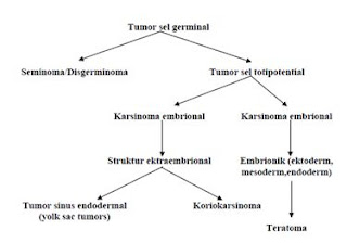
Teratomas are germ cell tumors are generally composed of multiple cell types derived from one or more of the three germ layers of endoderm, mesoderm and ectoderm. Inconsistent nomenclature is often confused about the various subtypes of teratoma. Teratomas derived from the Greek meaning of the terrace monster, which was first described by Virchow in the first edition of his book published in 1863. Teratoma is divided into three categories: mature teratoma (benign), immature teratoma and teratoma with differentiation monodermal particular degrees depending on the quantity of immature tissue showed the potential for malignancy. Cystic teratomas are generally benign and malignant solid is. Teratomas vary from a benign form of the cystic lesions of well differentiated (mature) to form a solid and malignant (immature). Immature teratomas are malignant germ cell tumor to the three most common after disgerminoma and endodermal sinus tumor. In addition, there also have a specific component (usually squamous) who experienced malignant transformation, but are rarely found.

In 1831, Leblanc created the term dermoid cyst in the literature of veterinary medicine when he succeeded in removing skin lesions at the base of the skull resembles a horse. Both teratomas and dermoid, a term still used and are often used interchangeably. Implications of these elements behawa beginning to resemble the skin with a complementary structure composed of dermoid, whereas teratoma has no such limit. Dermoid now known as the trigeminal and contain various types of tissue.

Friday, 6 April 2012

PLASTIC / Oncologic Surgery SERIES: THE DISTINGUISHING Bell's palsy with Iatrogenic paralise of the facial nerve (etiology, SIGN-symptoms, DIAGNOSIS AND MANAGEMENT)


PLASTIC / Oncologic Surgery SERIES: THE DISTINGUISHING Bell's palsy PARALISE iatrogenic facial nerve (etiology, SIGN-symptoms, DIAGNOSIS AND MANAGEMENT)


The term Bell's palsy is a peripheral facial nerve paresis of unknown cause (idiopathic) and acute. Many are mixed up between Bell's palsy with peripheral facial nerve paresis other unknown causes.
Bell's palsy is found, usually people with facial paralysis learn from friends or family or at the mirror or brush teeth / rinsing. When the patient realizes that he is paralyzed on her face, then he began to feel fear, shame, low self-esteem, cosmetic and sometimes disturbing soul depressed, especially in women and in patients who have a profession that requires him to appear in public. Often the question arises in his heart, if his face could go back to normal or not.

from wikipedia picture
 from freedictionary.com

Thursday, 5 April 2012

SIGN, SYMPTOMS, DIAGNOSIS, AND MANAGEMENT SPINA BIFIDA


CHAPTER I
INTRODUCTION

The Spina bifida is a congenital defect of the posterior arch of the spine due to failure of closure of the neural elements of the spinal canal in the early development of embryos. (1,2,3)
In the early stages of neural plate formation of a gap is formed which then form the neural neural tube. Pipe is then a neural network of the brain and spinal cord. While in the womb, the tissue that forms neural tube does not close or not closed properly. This led to the opening in the vertebrae, which surrounds and protects the spinal cord. The process of neural tube closure takes place during the fourth week of embryonic life, and usually before a woman knows her pregnancy and ended. Neuralisasi process began at the dorsal midline and continues to sefal and caudal direction. The most recent closure occurred at the posterior end of the day-to-28. (3.4)
Sometimes it does not close the neural groove, this is because the error induced by chorda spinalis which lies beneath or under the influence of environmental teratogenic factors neuroepitel cells. Neural networks in this case remains open to the outside world. Disruption of this process causes neural tube defects which are then classified as disrafisme. Disrafisme divided into two cranial and spinal. (3,4)
Disrafisme spinal / myelodysplasia is a congenital anomaly of the spinal fusion caused by the failure of the structures at the midline. When the lesions confined to the bones (arch) whether one or more posterior levels, the disorder is referred to as spina bifida. (4, 5, 6)
If the neural elements involved it will cause paralysis and loss of sensation and sphincter disorders. The degree and localization of defects that occur vary. Light on the circumstances which may be found only a failure of fusion of one or more of the posterior arch of the vertebra at the lumbosacral region. Sometimes this disorder does not cause significant clinical symptoms. (1, 2,4,7)
Often the event of a defect in the posterior arch will give rise to disorders of the skin surface of the cover, which looks like a dimple, tuft of hair, skin or sinus fat mass.
Spina bifida can be classified into two types namely, spina bifida occult spina bifida and aperta (cystica). (1)


CHAPTER II
REVIEW REFERENCES

DEFINITION 
Spina bifida is a developmental anomaly characterized by defective closure of the bones in the spinal cord sheath so that the lining of the spinal cord and meninges may protrude (spina bifida cystica), or does not protrude (spina bifida occulta) .(2)
Several hypotheses of spina bifida include: (4)
1. Cessation of neural tube formation process as a particular cause
2. Excess pressure in the central canal of the newly formed rupture surface, causing neural tube
3. The damage to the walls of the newly formed neural tube as a cause.

Protrusion of the spinal cord and meningens cause damage to the spinal cord and nerves, resulting in decreased or impaired function of the body parts supplied by these nerves, or at the bottom. Symptoms depend on the anatomical location of spina bifida. Most occur in the lower back, the lumbar or sacral region, due to the closure of the vertebrae in place at the end of this section.
         

                                                                 Figure 1. Spina Bifida

Wednesday, 4 April 2012

TYPE OF NEUROFIBROMATOSIS OR VON RECKLINGHAUSEN'S DISEASE ( BENIGN DISORDER BUT CREEPY DISEASE ) : DEFINITION, ETIOLOGY,SIGN , SYMPTONS ,PROGNOSiS, DIAGNOSIS AND MANAGEMENT


CHAPTER I
INTRODUCTION

Neurofibromatosis is an autosomal dominant disorder that affects the bone, nervous system, soft tissue, and skin. At least 8 different clinical phenotypes of neurofibromatosis have been identified that are associated with at least two genetic disorders. Clinical manifestations increase over time. Neurological and developmental problems toward malignancy might come afterwards.

Neurofibromatosis type 1 have varying phenotypic expression including dermatologic manifestations. Some patients may have a particular expression of the skin, while others may have life-threatening complications.

A neurocutaneous condition, neurofibromatosis may involve almost any organ system. Thus, the signs and symptoms can vary widely demonstrated. Two major subtypes exist: neurofibromatosis type 1, also known as von Recklinghausen neurofibromatosis, which is the most common subtype and is referred to as the neurofibromatosis, and neurofibromatosis type 2, the so-called central neurofibromatosis is a genetic disorder associated with bilateral vestibular schwannomas multisystem, schwannomas spinal cord, meningiomas, gliomas, and juvenile cataracts, skin with lack of features.


CHAPTER II
REVIEW REFERENCES

II. A. DEFINITION
Neurofibromatosis is a genetic disorder that interferes with the growth of cells in the nervous system, causing tumors to form in nerve tissue. These tumors can occur anywhere in the nervous system, including the brain, spinal cord, and nerves of large and small. Neurofibromatosis is usually diagnosed in childhood or early adulthood.

Tuesday, 27 March 2012

MALIGNANT SKIN TUMOR / SKIN CANCER AT AGLANCE ( etiology,sign, symptoms,diagnosis and management of Basal Cell Carcinoma, Squamous Cell Carcinoma,and Malignant Melanoma )

 Malignant SKIN TUMOR AT AGLANCE
  ( Basal Cell Carcinoma, Squamous Cell Carcinoma, Malignant Melanoma )

Definition
Malignant skin tumor is a disease characterized by the growth of skin cells that are not controlled, can damage surrounding tissue and can spread to other parts of the body. Because the skin consists of several types of cells, the skin cancer also vary according to cell type affected.

Epidemiology
Adult skin cancer tend to have increased in number especially in the Americas, Australia and Britain. Based on several studies, those white people are more likely to suffer this type of skin cancer. It is predicted as a result of their frequent exposure (lots of exposure) sunlight. In Indonesia people with skin cancer is fairly small compared to the third-countries, however, skin cancer needs to be understood because in addition to causing defects (damage the appearance) is also at an advanced stage can be fatal to the patient (1).
Malignant skin tumor types that are found throughout the world are basal cell carcinoma (basalioma), squamous cell carcinoma, a relatively non melanoma and malignant melanoma. Basal cell carcinoma is the most common. In America, about 800,000 people suffering this cancer every year. 75% of skin cancer are basal cell kanser. Squamous cell carcinomas are found to be what the 200,000 Americans each year. Melanoma is the most rare but cause the most deaths. According to WHO, as many as 160,000 people  suffering melanoma each year and as many as 48 000 deaths are reported each year (3).

Skin Embryology (4)
The skin has a double origin:
(A) the superficial layer (epidermis), formed from the surface ectoderm.
(B) inner layer (dermis), derived from the underlying mesenchyme.

A. Epidermis
At first, the embryo diilapisi by a single layer of ectoderm cells (Fig. 20.1A). At the beginning of the second month, the epithelium is split and formed a flat layer of cells, or epitrikium periderm, on its surface (Figure 20.1B). In the subsequent cell proliferation in the basal layer, forming a third zone (intermediate zone) (Figure 20.1C). Finally, at the end of the fourth month, the epidermis obtain definitive arrangement, and can be recognized four layers (Fig. 20.1D)


Wednesday, 21 March 2012

NEURO SURGRY SERIES : ETIOLOGY,SIGN , SYMPTON ,DIAGNOSIS AND MANAGEMENT CEREBRAL ABSCESS

INTRODUCTION
         
The incidence of cerebral abscess case /100.000 population is estimated at 1 per year. This incidence decreased after 1950 in line with the increasingly widespread use of antibiotics. Comparison of prevalence between men and women are 3:1.
75-90% is a solitary abscess, of which 35-45% are located in the frontal lobe, temporal lobe at 30-40%, 15-20% in the parietal lobe, and 15% in the occipital, cerebellum and brain stem.

DEFINITION

Brain abscess is an infection process by which localized pus between brain tissue caused by a wide variety of bacteria, fungus and protozoa. Brain abscess were present in all ages. Most at the age of the second decade of life, between 20-50 years.Comparison between male patients with female is 3:1.

Etiology and predisposing

Thursday, 8 March 2012

Etiology,sign, symptoms,Diagnosis and management Benign Prostat Hyperplastic (hyperplasia / hypertrophy)

Introduction
Enlargement of the prostate gland has a significant morbidity in a population of elderly men. The symptoms are a common complaint in the field of urologic surgery.
Prostate hyperplasia is one of the major health problems for men over the age of 50 years and was instrumental in the decline in quality of life. A study says that one third of men aged between 50 and 79 years of experience prostatic hyperplasia.
Exact prevalence in Indonesia is not yet known but is estimated based on foreign literature since age 50 years 20% -30% of patients will require treatment for prostatic hyperplasia. Which obviously depends on the prevalence of child bearing age. Actually, the changes towards the enlargement of the prostate is started early, beginning in mikroskopoik changes which then manifests into macroscopic abnormalities (enlarged gland) and then a new clinical symptoms.
Based on the autopsy rate is microscopic changes in the prostate can be found at the age of 30-40 years. When these microscopic changes constantly evolving there will be changes anatomic pathology. In men aged 50 years and the number of events around 50%, and at the age of 80 years about 80%. Approximately 50% of the numbers mentioned above will cause the symptoms and clinical signs.
The existence of this hyperplasia would lead to urinary tract obstruction and to overcome this obstruction can be done in various ways ranging from the most minor of action that is a conservative (non operative) to the most severe action that is operating.
I. ANATOMY AND PHYSIOLOGICAL
Under normal circumstances the prostate is approximately the size of a walnut. Normal prostate weight in adults is ± 20 grams. Located around the prostatic urethra and bladder neck as well as between the urogenital diaphragm. The apex of the prostate is located on the external urethral sphincter of the bladder. In the anterior symphysis pubis adjacent to but separated by a cavity in the extraperitoneal fat retopubis (Retzius cavity). In the posterior prostatic fascia is separated from the rectum by denonvilliers.


Thursday, 23 February 2012

Hydrocele and hernia at a Glance (etiology,sign, symptoms,diagnosis and management)


A.      ANATOMY
Generally hernia is a protussion the content of the cavity through  defect or the weakness of the cavity wall in question. On abdominal hernia, abdomenal content protunding  through a defect or weakness of the musculo-aponeurotik layer of the abdominal wall. Based on the occurance, congenital hernia or hernia are divided into congenital and acquired hernia. Hernia are named according to its location, eg diaphragm, inguinal, umbilical and femoral


By their very nature, can be called a hernia hernia when the hernia contents can reponibel jeluar entry. Out when standing or straining, and came back when sleeping or pushed into the stomach. When the contents of the bag can not be repositioned back into the abdominal cavity, called a hernia hernia ireponibel. This is usually caused by adhesions in the peritoneal pouch bag hernia. This is called a hernia hernia accreta. There is no pain or signs of intestinal obstruction.

Inkarserata called hernia or hernia strangulate when it squashed by the hernia ring so that the bag can not be caught and returned to the abdominal cavity. As a result, frequent passage or vascular disorders. Inkarserata hernia clinically more intended for hernia ireponibel with passage disturbance, whereas vascular disorder known as Strangulated hernia.
External hernia is an abnormal protrusion of intra-abdominal organs through the abdominal wall defect in the fascia. Hernia which often happens is inguinal, femoral, umbilical, and paraumbilikal.

Inguinal hernia is the protrusion viscus (organ) from the peritoneal cavity into the inguinal canal.

All hernia occurs through slit weak or potential weakness in the abdominal wall that is triggered by an increase in intra-abdominal pressure that repeated or continuous.


Seventy-five percent of all abdominal hernias occur in the inguinal (groin). Others may occur in the umbilicus (navel) or other abdominal area. Inguinal hernias are divided into two, namely the medial inguinal hernia and inguinal hernia lateralis. If the bag reaches the lateral inguinal hernia scrotum (testicles), called a hernia hernia skrotalis. Lateral inguinal hernia occurs more frequently than the medial inguinal hernia with a ratio of 2:1, and among men it was 7-fold more frequently affected than women. The more we age, the greater the possibility of a hernia. This is influenced by the strength of abdominal muscles that have been weakening.

Hernias that arise in the crease and the inguinal hernia abdominokrural is arising below the fold is a femoral hernia. Inguinal canal is an oblique line passing through the lower anterior abdominal wall. This channel allows the structures to pass to and from the testis to the abdomen in the male. In women, the channel is crossed by ligaments rotundum uteri, magi from the uterus to the labium. In addition, the channel is bypassed Ilioinguinalis nerve in both sexes.
            The length of the inguinal canal in adults is approximately 4 cm, formed from the profundus inguinal annulus / annulus inguinali internal to the superficial / external. Inguinal canal is located parallel to and just above the inguinal ligament. In neonates, the internal inguinal annulus is located almost directly posterior to the external inguinal annulus so that the inguinal canal is very short at this age. Then, the internal annulus moves toward the lateral growth effect.
            Internal inguinal annulus is an oval hole in the fascia transversalis, located about 3 cm above the inguinal ligament, midway between the Messiah and symphisis pubis.Medial to the internal annulus there is av. inferior epigastric. Edge of the annulus is the origin of the internal spermatica fascia in the male or the inner wrapper rotundum uteri rotundum ligaments in women.
            Externa is an annular shaped defect in the inguinal triangle (Hesselbach's triangle) on the aponeurosis. Obliquus externus abdominis and essentially formed by the crista pubica. Edge of the annulus is the origin of the fascia spermatica externa. Lateral boundary is the inferior epigastric artery, the medial border of the m. rectus abdominis lateral part, and the inferior border of the inguinal ligament.
            Inguinal canal is formed on the wall of the anterior, posterior, superior, and inferior.Anterior wall formed by the aponeurosis m. Obliquus externus abdominis is amplified at 1/3 by the lateral fibers of m. Obliquus internus abdominis. The entire length of the posterior wall of the inguinal canal is formed by the fascia transversalis tendon reinforced cojoint in 1/3 medial. Cojoint tendon is the tendon insertion combined m. Obliquus internus abdominis and m. transversus abdominis is attached to the crista pubica and linea pectinea. Basic or inferior wall of the inguinal canal is formed by the inguinal ligament, while the roof is formed by m. Obliquus abdominis internus abdominis and m.transversus.

Figure 1. Hesselbach's triangle
Inguinal hernias can be directly (direct) and can also indirectly (indirect). Indirect inguinal hernia bag of walking through the deep inguinal ring, lateral to the inferior epigastric vessels, and finally towards the scrotum. Pockets of directors inguinal hernia protruding directly through the base of the inguinal canal, medial to the inferior epigastric vessels, and rarely go down towards the scrotum. Femoral hernia is almost always seen as a mass irredusibel, although pockets more kososng, because fat dam of femoral canal lymph nodes around the bag. Single, enlarged lymph nodes can mimic femoral hernia very quickly.
Indirect hernia bag is actually a process vaginalis persistently dilated. Hernia was walking through the deep inguinal ring and follow the sheath into the scrotum. At the deep ring, bag filling anterolateral side of the cord. Properitoneal fat pockets often associated with indirect and is known as a lipoma of the cord, although the fat is not a tumor.
Retroperitoneal organs such as the sigmoid colon, cecum and ureter can be slipped into a pocket indirect. In the bag, these organs become part of the pouch wall and are susceptible to injury during repair.
Inguinal hernia pouch directors come from the bottom of the inguinal canal, the Hesselbach triangle; protruding hernia bag directly and does not contain obliqus externus muscle aponeurosis. Only in rare circumstances, the hernia is so intense that it pushes out through the annulus superficial and descend into the scrotum. The bladder is often a component of the bag is empty hernia director.
Bags from the femoral hernia femoral canal through a defect on the medial side of the femoral sheath (femoral sheath). Femoral canal contains one or two lymph nodes, which are scattered called Cloquet. Lymph-nodes are pushed out of the femoral canal by a protrusion of peritoneal and often form a palpable mass.


Phylloid Tumor (Mamae) / Cystosarcoma phylloides Mamae ( etiology,sign, symptoms,diagnosis and management)


                                                                     CHAPTER I
                                                                INTRODUCTION

It is a benign neoplasm that is locally infiltrate and possibly malignant (10-15%). Used to be called Cystosarcoma phyllodes are benign tumors that occur almost exclusively on women's breasts. Its name comes from the Greek word sarcoma, which means the fleshy tumor, and phyllo, meaning leaf. So called because the tumors display characteristics of a large, malignant sarcomas, such as display-leaves when cut, and the epithelium, cyst-like space when viewed histologically. Because most of the tumor was benign tumor is also called filoides.
Although metastatic tumors are not benign, but they have a tendency to grow locally aggressive and recurrent. Similar to sarcoma, malignant tumor hematogenous metastasis. Filoides tumor pathological picture is not always predict the clinical behavior of neoplasms, hence in some cases there is uncertainty about the classification level of the lesion.
The characteristics of malignant tumor filoides are as follows:
· Recurrent malignant tumors are more aggressive look than the original tumor.
· Pulmonary metastasis is the place most frequently, followed by bone, heart, and liver.
· The symptoms for metastatic involvement can occur immediately, until a few months and no later than 12 years after initial therapy.
· Most patients with metastases died within 3 years of initial therapy.
· There is no treatment for systemic metastases occurred.
· Roughly 30% of patients with malignant tumors filoides died of the disease.

            Filoides tumor etiology is not known, it is estimated associated with fibroadenoma in some cases, because patients may possibly have a second histological lesions in the same tumor. However, if the tumor develops filoides of developing fibroadenoma or both together, or whether the tumor can arise de novo filoides, is still unclear
This tumor has a smooth texture, sharply bounded and usually move freely. This tumor is relatively large tumor, with an average size of 5 cm. However, lesions> 30 cm have been reported.
Haagensen reported roughly one for every 40 filodes tumor fibroadenoma. Distribution of age according to Haagensen majority occur between the ages of 35 and 55 years.Bilateral tumors are very rare and rarely also occur in patients under age 20, first appeared benign react.
Mammography and ultrasound examination (Figure II.03) are generally important in the diagnosis of breast lesions, but they are notoriously unreliable in differentiating benign tumor from filoides malignant form of the condition or of fibroadenoma. Thus, the findings on imaging studies is not a definite diagnosis. Open excision biopsy of breast lesions smaller or incisional biopsy for larger lesions is a surefire method for diagnosing tumor filoides.

 image 03. Mammography image of the tumor filoides